In case of developmental defects (so-called malformations), which is pectus excavatum, the only means of correcting the defect is surgical intervention.
Physical therapy, massage and sports They help only with deformations resulting from external mechanical influence.
Vitamin and hormonal preparations can help if the deformity is caused by metabolic disorders or general hormonal development
However, this does not mean that exercise is excluded as an ineffective method. Moreover, sports, massage, and gymnastics are a necessary (but not sufficient!) attribute of treatment; they are necessary for the patient’s complete recovery.
The use of the Nuss method in the correction of pectus excavatum allowed the patient to engage in sports during the recovery period, which made the correction of deformities even more effective. Therapeutic exercise and massage are indicated during the period when the plate is installed on the patient: This promotes muscle development, as well as the formation of correct posture. Sufficient attention to physical exercises allows you to achieve excellent results in the correction of pectus excavatum.
Exercises for chest deformities
According to statistics, for every 1000 children, 6-8 have congenital chest deformities. The cause of chest deformities is considered to be an abnormal growth of cartilage between the ribs and the sternum, which bends the sternum inward or outward. In the first case, the anomaly is called a funnel-shaped deformity (Pectus Excavatum), in the second - a keeled deformity (Pectus Carinatum). In some cases, these deformities can be effectively treated using non-invasive methods, including physical therapy.
As the muscles are trained, the chest deformity can be reduced, or at least not worsened. In addition, intensive pumping, especially in the torso area, allows you to increase muscle mass and create a good cosmetic effect - the curvature becomes less noticeable.
All physical exercises are aimed at solving the following problems:
- Increase mobility and flexibility of the spine and chest.
- Stretch all shortened structures.
- Strengthen the muscles for a more amplitude movement of the chest.
- Restore normal posture.
Exercises mobilize joint mobility, stretch soft fabrics around the sternum so that they do not interfere with the movement of the chest. In addition to the mobility of the joint-ligamentous apparatus, muscle tone improves.
Suggested Exercise Program
Stretching and mobilization exercises
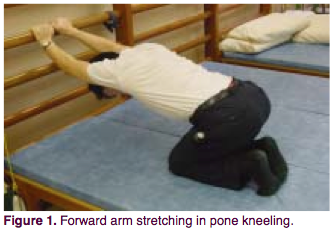
The patient is positioned on bent knees, arms extended forward. Holding the wall bars at a height of 50-80 cm, slowly lower top part body, and move your shoulder blades towards the floor. You should feel a stretch in the front armpit and shoulder. Hold for 8 seconds (you can breathe deeply, increasing the chest stretch), then relax. Repeat 20 times. Such approaches should be done 4 times a day.
The purpose of the exercise is to stretch all the anterior muscles of the chest, especially the pectoralis major muscle.
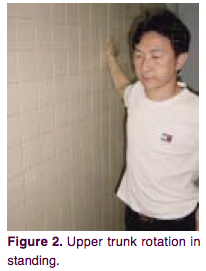
The patient stands sideways to the wall. Place the hand closest to the wall on the wall at a level slightly above the shoulder. Rotate your pelvis in the opposite direction, without lifting your hand from the wall. The stretch should be felt in the front of the shoulder and upper chest. Hold for 8 seconds. Then you can relax and return to the starting position. Rest and continue with the other hand. You need to do 4 sets of 20 repetitions per day.
This exercise gives the greatest range of motion of the thoracic vertebrae, allowing you to stretch the ligaments, muscles and joints in the sternum area.
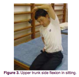
The patient is in a sitting position. Side bends are performed. When bending to each side, the opposite arm reaches overhead in the direction of the tilt. You should feel a stretch on the opposite side of your torso. Hold the position for 8 seconds (during this time you can breathe deeply), return to the starting position. Repeat exercises 20 times 4 times a day. The purpose of the exercise is the same as in the second exercise.
Strength exercises
4, 5. Exercise with weights in a lying position.
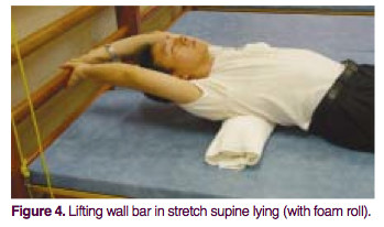
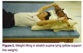
The patient is positioned in a supine position. You need to place a polyurethane foam roller about 10 cm in diameter under your back. If using a roller is too painful, just lie flat. Stretch your arms back behind your head and grab the bar of the wall bars or some heavy load at a distance of 10 cm from the ground. After this, take a deep breath and try to apply maximum effort, as if lifting a wall bar or a load. Hold the position for 8 seconds, then relax. 10 such repetitions constitute one series. You need to do 3 series 4 times a day.
Purpose of the exercise:
In this position, when the arms are fixed, the anterior chest wall rises mainly due to the pectoral muscle. Maximum tension when lifting force is applied causes the surrounding muscles involved in the breathing process to mobilize. A cushion placed under the back helps to expand and straighten the thoracic spine, correcting thoracic kyphosis.
6. Upper body stretch while lying on your stomach.
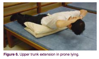
The patient lies on his stomach. You can place 1-2 pillows under your stomach. Hands behind your head. The legs can be fixed on the wall bars. Inhale deeply and raise your upper body, keeping your hands behind your head. Bend over. Hold this position for 8 seconds, then relax. 10 such repetitions constitute one series. You need to do 3 series 4 times a day.
The purpose of the exercise is to strengthen upper muscles back, balance muscle strength. This helps prevent thoracic kyphosis and maintain beautiful posture.
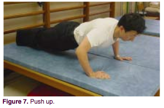
The patient lies on his stomach and begins to do push-ups. The easiest option is to lift only the torso, the more difficult is to lift the whole body, and the most advanced option is to clap your hands in the upper phase of the push-up, lifting your hands off the floor. Start with the first option. If it turns out easy, move on to the next one. One series is 10 repetitions. After each episode there is a rest. In total, you need to do 30 repetitions 4 times a day.
The purpose of the exercise is to generally strengthen the chest muscles. With intense loads, bone mineralization also improves; power pumping stimulates the formation of a beautiful silhouette.
8. Move your arms up and down along different sides of the body.
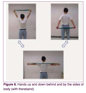
The patient is in a sitting or standing position. Arms extended. In your hands you need to hold the ends of an elastic band or any other elastic cord, its elasticity allowing you to do a maximum of 10 repetitions of the exercise, no more. Slowly move your arms back, lowering them to the level of your buttocks. Hold for 3 seconds. Then also slowly return your arms over the top to their original position. Repeat 10 times (1 series). After that, rest and do 2 more episodes. As a result, you need to do 30 repetitions 4 times a day.
The exercise serves to strengthen the neck, shoulders, upper back, pectoral muscles. It can be considered as a stabilizing exercise for the upper chest.
Chest deformity is a change in the shape of the musculoskeletal frame of the upper body. There are two main types of chest deformity in children: pectus excavatum and pectus carinatum.
What is associated with chest deformation in children, and what should parents do in case of such a diagnosis?
The health consequences associated with chest deformities in children depend on the type of deformity and its severity.
Funnel chest deformity in children is manifested in the retraction of the costal cartilages, resulting in the formation of a funnel, or depression, in the center of the chest.
There are 4 degrees of funnel chest deformity in children, depending on the depth of the funnel. With degree I deformation (a depression of no more than 2 cm), the child may not feel any symptoms of the disease at all. At higher degrees of deformation, the child may experience difficulty breathing, shortness of breath, and some disturbances in functioning. internal organs due to their compression.
With keeled chest deformity in children, the sternum protrudes forward in the form of a keel, to which the ribs are attached at right angles. This deformity is often only a cosmetic defect. If carinatum deformity has a pronounced character, this can lead to problems in the functioning of the lungs, heart and other internal organs due to a violation of their relative position. In this case, it is necessary to conduct an examination and find out the features of the location and functioning of the child’s internal organs.
What can cause chest deformation in children?
Chest deformity in children is most often a congenital disease and is formed during the prenatal period, when the child is in the mother’s womb. Scientists have not yet found an exact answer why the baby’s chest is deformed. It is only known that the likelihood of this defect increasing when:
Negative heredity (the presence of this disease in the medical history of the child’s mother or father or their closest relatives);
exposure to teratogenic factors (negative factors affecting the pregnant woman and the fetus and causing disturbances in its development without affecting hereditary structures). These factors include the transfer expectant mother infectious diseases, taking antibiotics and other chemicals, contact with radiation, etc.
That is, expectant mothers need to follow standard recommendations: take care of themselves, do not contact sick people, use with caution. medicines, etc.
As for acquired deformation of the chest in children, it can be caused by serious illnesses suffered by the child (rickets, scoliosis, pulmonary diseases, etc.) and injuries to the upper part of the body.
How is chest deformity corrected in children?
Mild chest deformities in children are treated conservatively, without surgery. It consists of carrying out physiotherapeutic procedures, massage, therapeutic exercises and, if necessary, wearing the child with special compression devices, orthoses and dynamic compression systems.
In more serious cases, children are prescribed surgery to correct the shape of the chest. Previously, it was believed that the younger the child being operated on, the better, since the ability of children's tissue to regenerate is much higher than that of a teenager or adult. Therefore, operations to correct the shape of the chest were performed on children in preschool age. However, now most doctors agree that early surgical correction of the chest shape can lead to abnormal growth of the ribs, recurrence of the disease and the need for repeated surgery. Therefore, surgeons recommend performing the operation no earlier than 10-12 years for boys and 12-13 years for girls.
Breathing exercises and physical therapy for chest deformities in children
The first thing you need to do if you notice a chest deformity in a child is to consult a doctor (orthopedic surgeon or more specialized specialist). If a specialist confirms that the defect does not pose a risk to the child’s health, parents can combat the deformation of the child’s chest on their own, namely, engage in breathing exercises and physical therapy with the child. These methods cannot completely correct the defect, but can slow down its development.
Breathing exercises for chest deformation in children help correct the shape of the musculoskeletal frame, and also normalize the functioning of the heart and lungs. Before doing breathing exercises with your child, you should check with your doctor to see if there are any contraindications to these exercises.
Breathing exercises
1. Holding your breath. Stand straight, feet shoulder-width apart. Take a deep breath and hold your breath for as long as possible. Then exhale sharply through your mouth. Repeat 5-10 times.
2. Upper breathing. Can be performed both standing and sitting. Inhale slowly and deeply, making sure that your stomach remains still and your chest rises. Exhale sharply through your mouth, repeat 5-10 times.
3. Expansion of the chest. Stand up straight, take a deep breath, clench your fists and extend your arms in front of you at shoulder level. With a quick movement, move your arms back and smoothly return to the starting position. Repeat several times and exhale sharply through your mouth. During the exercise, the arm muscles should be very tense.
In addition to breathing exercises, it is very useful for children with chest deformation to perform exercises to develop the pectoral muscles: push-ups, pull-ups, exercises with dumbbells and an elastic gymnastic band. Strong muscles The chest will help slow down the deformation and even stop it; in addition, a developed muscular frame will visually correct the cosmetic defect and close the deformed chest.
Children with deformed chests benefit greatly from swimming; this sport helps develop the pectoral muscles and lungs and has very few contraindications. Volleyball, basketball and rowing are also often recommended for this disease, especially if the child shows interest in them.
Mild chest deformation in children usually does not affect their health, especially if parents take measures to correct the defect: they do breathing exercises with the child, teach him to play sports. And even if the degree of deformation is high, medicine suggests effective ways complete elimination of the defect, ranging from high-tech compression devices to modern operations with minimal intervention.
Grow up healthy!
THERAPEUTIC PHYSICAL CULTURE
WITH FUNNEAL CHEST.
Congenital pectus excavatum in children is one of the most severe orthopedic diseases.
There is no consensus among pediatric surgeons about the causes of this disease.
The opinions of a number of researchers about rickets as the cause of the development of pectus excavatum are not currently confirmed.
N.I. Kondrashin (1973) believes that breast deformation is a consequence of dyschondroplasia of the costal cartilages and sternum. Many researchers share this opinion.
In children, the chest with a funnel-shaped deformity has a characteristic shape. It is flattened, the sternovertebral distance is reduced, the ribs take on an incorrect inclined or oblique position, and the spigastric angle is sharpened.
There are three degrees of chest deformation.
In grade I, the depth of the “funnel” does not exceed 2 cm, the heart is not displaced. In degree II, the “funnel” is deeper, 3-4 cm. In degree III, the depth of the “funnel” is more than 4 cm, the heart is displaced by more than 3 cm.
Children with this disease tend to be significantly delayed in physical development. They are noticeably underweight. Posture is disrupted and the spine is bent. Physical performance is clearly reduced.
A deformed chest creates unfavorable conditions for the functioning of the thoracic organs. First of all, the function of external respiration is impaired. In patients, the drainage function of the bronchi becomes difficult, pulmonary ventilation decreases, and the chest circumference and excursion are reduced compared to the age norm.
Redox processes clearly change, the acid-base state of the blood is disrupted, mainly towards the development of respiratory alkalosis and metabolic acidosis. Under these conditions, oxygen deficiency develops, which significantly affects the functioning of the main vital organs. The cardiovascular system functions under conditions of mechanical compression and displacement of the heart. All this leads to a significant decrease in the reserve capacity of the cardiovascular and respiratory systems. Sinus tachyarrhythmia develops. With severe deformities, blood pressure may change and venous pressure may increase. There is a certain tendency to vegetative-vascular dystonia.
Children with this disease are lethargic, irritable, get tired quickly, do not recover well from physical activity, and often complain of pain in the heart area and heaviness in the chest.
Surgical treatment is required for funnel chest deformities with a tendency to progress, accompanied by a significant decrease in the reserve capacity of the cardiovascular and respiratory systems. An important indication for thoracoplasty is the presence of a cosmetic defect.
In preparing the patient for surgery important has medicinal physical culture, the main objectives of which are a general tonic effect, strengthening weakened muscles, especially the pectoralis major and minor muscles and back muscles, correcting posture, improving respiratory and circulatory function, teaching early physical exercises postoperative period.
Therapeutic physical culture of the preoperative period is carried out in the form of hygienic and therapeutic exercises. The total load during classes is determined depending on the age of the children, the degree of deformation and the functionality of the cardiovascular and respiratory systems. Therapeutic gymnastics classes are carried out from the starting position, standing and lying down, using equipment (clubs, hoops, gymnastic sticks, balls, medicine balls, rubber bandages, etc.). In the construction of classes, they adhere to generally accepted forms, observing the principles of consistency and gradualism. For children of preschool and primary school age, physical exercises of an imitation nature are included, and the game method is used.
We present an approximate complex of therapeutic exercises for school-age children before surgery for funnel chest (II degree).
1. Construction, alignment of posture in front of the mirror.
2. Walking at different paces and with varying degrees of difficulty for 1 1/2-2 minutes.
3. Main stance - touching the plane with your back, buttocks and heels. Sliding along the plane, raise your arms, palms turned forward, through the sides up, stretch, rising onto your toes. Repeat 4-5 times.
4. Standing, hands behind the castle. Bend over, bringing your shoulder blades closer together. Repeat 4- 5 times.
5. Standing with your feet shoulder-width apart. Rotate straight arms forward and backward while maintaining correct posture. Repeat in each direction 5-6 times.
6. Main stand. Raise your hands up - inhale, lower them down - exhale. Breathing through the nose. Repeat 4-5 times.
7. Standing, legs apart, gymnastic stick on the shoulder blades. Squats (maintain correct posture). Repeat 10 times.
8. Standing, legs apart, stick on shoulder blades. Rotate the body right and left. Repeat 5-6 times V each side.
9. Standing, legs apart, gymnastic stick in hands. Raise the stick up - inhale, lower the stick down - exhale. Repeat 4-5 times.
10. Walking with a stick on your shoulder blades. Raise your knees high and watch your posture. Duration 1-2 minutes.
11. Standing, legs apart, hands to shoulders. Tilt the body to the sides with raising your arms up. Repeat 4-5 times in each direction.
12. Standing, legs apart, medicine ball in hands. Lift the medicine ball up and throw it back. Repeat 4-5 times.
13. Main stand. Raise your arms up - inhale, relax your arms and alternately lower your hands, forearms and shoulders down - exhale. Repeat 3 4 times.
14. Standing with your back to the gymnastics wall, a rubber band is fixed to the crossbar at head level. The ends of the bandage are held outstretched to the side. Stretch the rubber bandage, bringing straight arms through the sides to the body. Repeat 3-4 times.
15. Lying on your back. Exercise "bicycle". Repeat 11-12 times.
16. Lying on your back. Static full breathing. Repeat 4-5 times.
17. Lying on your back. Flexion and extension of fingers and toes simultaneously and alternately. Repeat 10-12 times.
18. Lying on your back. Rotation in the wrist and ankle joints. Perform simultaneously and alternately. Repeat 10-12 times.
19. Lying on your stomach. Hands to shoulders. Raise the belt upper limbs» bringing the shoulder blades closer together. Repeat 6 times.
20. Standing on all fours. Raise alternately the right and left hand. Don't bend your back. Repeat 2-3 times with each hand.
21. Walking at a variable pace, 1-2 minutes.
22. Standing in the middle of a rubber bandage. The ends of the bandage are in the lowered hands. Stretch the rubber bandage by raising your arms straight forward. Repeat 3-4 times.
23. Ball game (volleyball, two-handed basket throws), 2-3 min.
24. Walk at a calm, slow pace, 1-2 minutes.
Considering the reduced reserve capacity of the cardiovascular and respiratory systems and increased fatigue in children, physical activity should be strictly regulated.
Therapeutic physical training in the postoperative period begins immediately the next day after surgery. At the first and early stage, it aims to prevent postoperative complications, primarily pneumonia, adapt the cardiovascular and respiratory systems to new operating conditions, combat muscle wasting and other negative consequences of prolonged hypokinesia.
The method of therapeutic physical culture depends on the technique of surgical intervention. At the Department of Pediatric Surgery of the 2nd Moscow State Medical Institute named after. N.I. Pirogov has been using the thoracoplasty technique developed by N.I. Kondrashin (1968) for a number of years. This radical operation eliminates the use of traction sutures and fixators. To achieve strong consolidation of the ribs and sternum in the position achieved through surgical correction, the child should remain for 30-45 days in a supine position without a pillow on a flat, hard board. Children are not allowed to stand up, sit down, or turn around.
In accordance with the proposed surgical treatment, we have developed the following method of therapeutic exercises.
The postoperative period is divided into early and late. The early postoperative period, in turn, is divided into periods “A” and “B”. Early period “A” is prescribed to the patient in the first 5 days after surgery. The early “B” period usually lasts 10-15 days after surgery. Therapeutic gymnastics is carried out 3 times a day.
An approximate complex of therapeutic exercises, applied on the 2nd day after thoracoplasty for congenital pectus excavatum deformity in children preschool age(Fig. 59.1-12).
All gymnastics are performed from the starting position lying on your back.
1. Foot massage-stroking. Repeat 6-8 times (Fig. 59.1).
2. Clenching and unclenching the fingers (“the peacock spread its tail”). Repeat 4-5 times (Fig. 59.2).
3. Clenching and unclenching your toes. Repeat 4-6 times (Fig. 59.3).
4. Rotation of the hands in the wrist joints. Repeat 4-6 times every day blowing side (“screwed the screws”) (Fig. 59.4).
5. Dorsal and plantar flexion at the ankle joints (“hello, goodbye”). Repeat 3-4 times (Fig. 59.5).
6. Static breathing exercise. Repeat 3-4 times (Fig. 59.6).
7. Flexion and extension of the arms at the elbow joints, without lifting the elbows and shoulders from the plane of the shield (“hammers clattered”). Repeat 3-4 times (Fig. 59.7).
8. Exhale several times rubber toy(rest) (Fig. 59.8).
9. Static tension of the quadriceps thigh muscles. Repeat 3-4 times (“move the kneecap”) (Fig. 59.9).
10. Turns the head to the sides. Repeat 2-3 times (“cross the street, look left, look right”) (Fig. 59.10).
11. Turn your arms palms up and inhale. Return to the starting position - exhale. Repeat 3-4 times (“show palms, hide palms”)! (Fig. 59.11).
12. Clenching and unclenching of the toes (“the hedgehog went hunting, the hedgehog | curled up into a ball”). Repeat 5-6 times (Fig. 59.12).
5 days after the operation, classes can be carried out according to the method of the early period “B”. During this period, as well as in the first days after surgery, classes are conducted from the starting position lying on your back. The total load gradually increases. Exercise should be included in your classes! for large muscle groups - flexion and extension of legs in co-| lazy.and hip joints, raising the pelvis.
The late postoperative period is divided into late “A” and late “B” periods.
Late period "A" usually starts on the 15th day and continues until the 30-40th day after surgery,
Therapeutic physical culture of the late period “A” is carried out in the form of hygienic and therapeutic gymnastics.
Hygienic gymnastics are carried out daily, immediately after children wake up. The purpose of hygienic gymnastics is a general tonic effect, facilitating the transition from sleep to wakefulness.
Therapeutic exercises are prescribed 2 times a day. Its main task is to prepare the child for a gradually increasing load, gradually train blood circulation, breathing, and strengthen skeletal muscles.
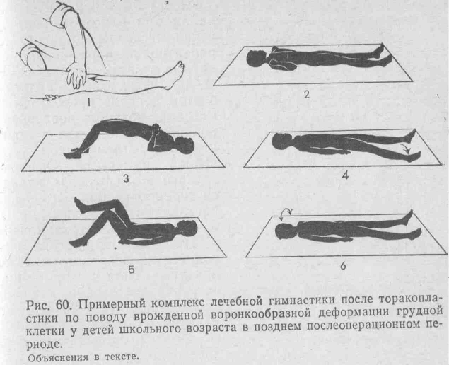
An approximate complex of therapeutic exercises for school-age children (on the 20th day after thoracoplasty) (Fig. 60.1-6)
Classes are conducted from the starting position lying on your back.
1. Foot massage-stroking. Repeat 8-10 times (Fig. 60.1).
2. Simultaneous squeezing of fingers and toes. Repeat 8-10 times.
3. Rotation of the arms in the wrist and ankle joints. Repeat 6-8 times (Fig. 60.2).
4. Static breathing exercises. Repeat 3-4 times.
5. Flexion and extension of the arms at the elbow joints without lifting the elbows from shield plane. Repeat 4-6 times.
6. Static tension of the quadriceps thigh muscles. Repeat 8-10 times.
7. Raising your arms to the sides - inhale, return to the starting position - exhale. Repeat 3-4 times without taking your hands off the plane of the shield.
8. Static tension in the gluteal and back muscles. Repeat 4-6 times.
9. Static breathing exercises. Repeat 3 -4times.
10. Raising the pelvis with support on the arms and heels bent at the elbows. Repeat 2 times. Rest pause 15 s (Fig. 60.3).
11. Raising your arms up - inhale, lowering your arms down through the sides - exhale. Repeat 3-4 times.
12. Moving the leg to the side without lifting the heel from the plane of the shield. Repeat 2-3 times with each leg (Fig. 60.4).
13. Static breathing exercises. Repeat 3-4 times.
14. Legs bent at the knee and hip joints - walking lying down. Repeat 10-12 times (Fig. 60.5).
15. Inflating the rubber ball until it is full. Rest 15 sec.
16. Alternate flexion and extension of the legs at the ankle joints. Repeat 8-10 times.
17. Turns the head to the right and left (Fig. 60.6).
18. Vigorously clench and unclench your fingers. Repeat 8-10 times.
19. Place your feet hip-width apart. Turn your toes out to the sides and return to the starting position. Repeat 3-4 times.
20. Turn your hands with your palms up - inhale, return to the starting position - exhale. Repeat 3-4 times.
After 5-6 weeks (after the operation, with a successful course of the postoperative period, patients are allowed to sit down and then stand up. Therapeutic physical training during this period is carried out according to the method of the late postoperative period “B”) in the form of morning hygienic and therapeutic exercises. The main task of this period is to train the patient under increasing loads and prepare for discharge from the hospital.
Therapeutic gymnastics is carried out from the initial position (lying, sitting and standing. The total load gradually increases and reaches 20-25 minutes. Classes according to the described method should be carried out daily until discharge home.
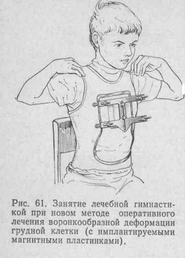
For recent years at the Faculty of Pediatric Surgery of the 2nd Moscow State Medical Institute named after. N. I. Pirogov Yu. F. Isakov, V. I. Geraskin, G. S. Vasiliev, C. C. Py-j Dakov (1979) developed new way surgical treatment! funnel chest deformity. In the chest! space is implanted with a magnetic plate, which allows|^ st with the help of a permanent powerful magnet, strengthened by n| surface of the chest, perform (gradual stretching of the chest! we to the position of the desired correction (Fig. 61). 1st
Such new method surgical intervention gave an effect | the ability to expand the motor mode in the postoperative period. In the absence of complications, patients are allowed to sit down on the 3-5th day, get up and walk on the 7-10th day. The corset is removed after about a month.
Therapeutic exercises in the postoperative period begin the next day after surgery.
The main task of the early postoperative period is the prevention of postoperative pneumonia, adaptation of the cardiovascular and respiratory systems to new operating conditions, and the fight against muscle atrophy and other negative consequences of physical inactivity.
The postoperative period is divided into early “A” and “B” periods. Period “A” is prescribed in the first 5-7 days after surgery. Patients are treated with therapeutic exercises from the starting position lying down. They include dynamic and static breathing exercises, which helps to cough up the contents of the bronchi, exercises for small, medium and large muscle groups. A mandatory exercise is the impact on the muscles of the pelvis and waist lower limbs. Execution rate physical exercise medium and slow. Rest pauses are included. The total duration of the lesson is 7-10 minutes. It is recommended to conduct classes at least 3 times throughout the day.
On the 7-10th day after surgery, when the patient is allowed to sit down, therapeutic exercises are prescribed according to the early postoperative period “B”. During this period, classes are conducted from the starting positions lying and sitting on a chair.
In a lying position, physical exercises for the upper and lower extremities are recommended (at a medium pace. A number of exercises are done with effort. Static and dynamic breathing exercises are used. The transition to a sitting position on a chair is carried out carefully, without sudden movements. From this position, careful bending in thoracic spine until the shoulder blades come together, alternately lifting the shoulders, slightly tilting the body to the sides, alternately and simultaneously raising the arms up (in the first days, this exercise is carried out with the help of all exercises from a sitting position, without jerking or sudden movements until painful ones appear). sensations. The duration of the lesson during this period increases to 10-15 minutes.
After 10-14 days, when the patient is allowed to stand on his feet, therapeutic exercises are carried out according to the method of the late postoperative period. During this period, a set of physical exercises is prescribed from the starting position lying, sitting and standing. Physical exercises are used with a greater (compared to the previous period) amplitude. They use gymnastic sticks, balls, hoops and other gymnastic equipment. Various physical exercises are selected for the upper extremities, bending in the thoracic spine until the shoulder blades come together, walking with a gymnastic stick on the shoulder blades, walking with the shoulder blades coming together, turns, bending to the sides, squats. Lying on your back - exercise "bicycle". During this period, classes can already be conducted in a small group method.
When constructing therapeutic gymnastics classes, the generally accepted principles of periodization are preserved.
The objectives of therapeutic exercises are to expand the functional capabilities of the cardiorespiratory system, adapt to the changed conditions of the function of the respiratory and circulatory system, and general training for increasing physical activity, strengthening skeletal muscles. The duration of the lesson increases to 15-20 minutes.
After removing the corset, the opportunity is used to more actively correct posture and strengthen skeletal muscles, especially the pectoralis minor and major muscles and back muscles. Exercises are used from the starting position lying on the stomach - “swallow”, exercises with resistance and with effort.
It is advisable to continue practicing therapeutic exercises in the physical therapy rooms of children's clinics.
6-10 months after the operation, if the course of the postoperative period is favorable, children can attend physical education classes at school in a special group.
In the future, according to indications, it is possible to transfer children to the preparatory and main groups.
Long-term results of complex treatment, monitored over several years, indicate the greater effectiveness of the proposed method: correction of posture, significant correction of chest deformity, a noticeable increase in physical performance, pronounced compensation for cardiorespiratory failure, which can also be explained by the training influence of therapeutic physical culture.
Heading:
With normal development, the chest in the first years of a child’s life is somewhat laterally compressed; its anterior-posterior size at this time is larger than the transverse one, the ribs extend from the spine almost horizontally. From the moment the child begins to take a vertical position - to sit, the relationship changes: the ribs in front take on an inclined position, the chest drops due to its heaviness, the heaviness of the organs located in the chest cavity, and the traction of the abdominal muscles. As the child grows, the anterior-posterior diameter of the chest begins to lag behind and, from the age of two, gradually gives way to the growth of the transverse diameter. Subsequently, within the limits of normal development, the rib cage in an adult gives two types: long, narrow and short, wide. Along with this, pathological forms are observed; some of them are secondary, such as changes in the chest due to various curvatures of the spine, others are independent. The latter include the following.
A flat chest is characterized by weakness of the dorsal and pectoral muscles; the weakness of the former leads to a forward tilt of the torso in its upper part, and the weakness of the latter leads to a lowering of the chest. In general, the flat chest is elongated and flattened; the shoulders protrude sharply forward, the shoulder blades are raised, the distance between them is increased. As a result of this change in the chest-non-cells, respiratory movements are difficult and the child requires special efforts to take a deep breath; usually it is limited to shallow breathing, which causes insufficient pulmonary ventilation, a decrease in blood oxidation and, as a result, leads to anemia and weakening of the overall development of the child.
A flat, narrow chest, due to its limited ability to expand, retards the development of the lungs, so their capacity is reduced; deep breathing due to weakness of the pectoral muscles, as already indicated, is difficult; heart with such chest reduced, its working conditions are unfavorable, since the suction activity of the chest is weakened. A flat chest also has a negative impact on the development and function of the abdominal organs. Reasons for the formation of a flat chest: rickets, narrowing of the first costal ring due to ossification of the costal cartilage, muscle weakness, especially spinal muscles, sitting for a long time at school under conditions unfavorable for development, poor living conditions, etc.
Prevention - proper physical education.
Treatment: corrective exercises to expand the chest and develop weak back muscles and shoulder girdle. Exercises on apparatus (bar, ladder), promoting passive stretching of the chest, raising the ribs and increasing their mobility; active exercises to strengthen and develop the chest muscles. Breathing exercises. General exercises for the development of the heart, lungs and all muscles. Ball games and other outdoor active games are widely recommended. Improving living conditions and school conditions.
Funnel chest can be congenital or acquired. The cause of the latter may be rickets or pressure on the chest from various devices in a particular profession. Funnel-shaped breasts are characterized by the fact that in the area of the base sternum and the xiphoid process there is a depression, which can be of different sizes. With an average degree of depression of any disorders of the thoracic and abdominal cavities does not happen; with a strong degree of development, there may be difficulty breathing and cardiac dysfunction.
Pectus excavatum is a deformity that is more persistent and difficult to treat than flat chest. The results of treatment are better the earlier it is started. It is recommended to apply air pressure to the depression in the chest bone from the inside - take a deep breath and exhale intensely (with a closed or narrowed glottis), and at the same time use your hands to compress the chest from the sides, since it is usually wider than normal in cross-section. In addition to general strengthening physical exercises, additional ones are given for the development of the chest in the sagittal direction. For example, raising your raised arms to the sides and back while taking a deep breath; circular movement of the arms extended forward, up, to the sides, back and down with deep breathing; mixed hangs on rings with circular rotation, etc. In case of very deep depression, accompanied by difficulty in the functioning of the organs of the thoracic cavity, surgery is indicated.
Keeled breast (chicken) is more common than funnel breast. The shape of the thorax is similar to the shape of a ship's keel or the shape of a bird's chest. Chicken breast develops due to rickets, when the softened costal walls are influenced by suction from the chest cavity and traction from the diaphragm; Secondarily, it can develop with rachitic and tuberculous kyphosis. The anterior-posterior diameter of the chest is increased, the transverse diameter is reduced, the sternum protrudes significantly forward; on the lateral surface of the chest, the ribs from the fourth to the eighth are concave, and the lower ones are convex and form belt-like protrusions with the overlying ones, defining the lateral flattenings even more prominently; The ribs approach the sternum at right angles. The collarbones are directed obliquely from front to back; often have angular bends. Traces of rickets in the form of rosary beads are visible on the ribs. This deformation negatively affects the activity of the heart and lungs; it retards the development of the latter, especially in the apices of the lungs, reducing gas exchange in them. But, in general, the prognosis for chicken breast is favorable, since over time the deformation gradually smoothes out, and mild forms are almost completely cured.
IN initial stage diseases are prescribed antirachitic drugs. For older children, corrective exercises are used: in a sitting position, raising the raised arms to the sides and back, while simultaneously squeezing the chest back, while taking a deep breath and exhaling; a similar exercise is performed lying on your chest. At the same time, general exercises are carefully done to strengthen the musculoskeletal system and internal organs, as well as breathing exercises.
- 1. Walking around the hall. Perform for 1-2 minutes.
- 2. Continuing walking, raise your arms up, take a breath, return to the standing position. - exhale. The duration of the exercise is 1-2 minutes.
- 3. I.p. - basic stance (legs together, arms down). Left leg back, arms up - inhale. Return to IP - exhale. Same with the right foot. The pace is slow. When performing the exercise, look forward. Perform 6-8 times with each leg.
- 4 I.p. - the same. Bend forward, arms to the sides - inhale, etc. - exhale. Repeat the exercise 6-8 times
- 5. I.p. - sitting on the floor, legs to the sides, hands behind you. Raise your pelvis from the floor, bend slightly, throwing your head back - inhale. Return to IP - exhale. Perform 4-6 times.
- 6. I.p. - lying on your back, arms along your body. Chest type of breathing. Repeat 3-4 times.
- 7. I.p. - Same. Alternating flexion of the feet. Will do 8-10 times.
- 8. I.p. - Same. Arms to the sides, bend your right leg, press your knee to your stomach, straighten your leg up, bend it again and lower it. Same thing with the other foot. Repeat 8-10 times with each leg.
- 9. I.p. - Same. Bend your arms to your shoulders, legs to the sides - inhale, return to the i.p. - exhale. Perform the exercise 8-10 times.
- 10. I.p. - Same. We perform the “Bicycle” exercise (twisting virtual bicycle pedals in the air). Repeat 8-10 times
- 11. I.p. - Same. Hands up, at the same time bending your legs, pressing them to your stomach at the knees - inhale, return to IP. - exhale. Perform 6-8 times.
- 12. I.p. - lying on your stomach, arms along your body. Arms to the sides forward, legs to the sides - inhale, return to the i.p. - exhale. Repeat 8-10 times.
- 13. I.p. - lying on your stomach, arms to your sides. Circular rotations with arms backwards. Repeat the exercise 8-10 times
- 14. I.p. - lying on your stomach, put your hands with a gymnastic stick forward. Bring the stick behind your back onto your shoulder blades - inhale. Return to i.p. - exhale. Maximum amplitude. Perform from 2 to 10 times.
- 15. I.p. - the same, arms to the sides, bend at 90 degrees. Bend your knees, put your arms forward, slightly raised. The pace is slow. Repeat 8-10 times.
- 16. I.p. - lying on your stomach, chin on the back of your hands, elbows to the sides. Raise your hands behind your head - inhale, return to standing position. - exhale. The pace is slow. Perform 4-6 times.
- 17. I.p. - lying on your stomach, grab your ankle with your hands, return to the position. The pace is slow. Number of repetitions 4-6 times.
- 18. Finish everything by walking around the hall for 1-2 minutes. The pace is slow.
 Exercise therapy for chronic cholecystitis. Semi-bed rest
 Exercise therapy for colitis accompanied by spastic constipation
 A set of therapeutic gymnastics exercises in the adaptation period for tuberculosis ( middle group)
 Exercise therapy on the second day after hernia repair for inguinal and femoral hernias
 Exercise therapy for chronic hepatitis. Free mode
 A complex of therapeutic exercises for tuberculosis used during the adaptation period (strong group)