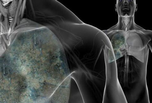
We often hear about lung cancer, but less often about an equally serious disease called COPD - chronic obstructive pulmonary disease. This term refers to a combination of two conditions: emphysema and chronic obstructive bronchitis. Both can be caused by lung damage from smoking or irritants such as asbestos. Whether you smoke, quit, or are simply unlucky, you can develop COPD as a result of damage to your lungs, which gradually limits your ability to inhale oxygen.
Signs you need to have your lungs checked
When your lungs are not functioning at their full capacity, symptoms may begin unnoticed and be difficult to recognize. Because COPD is a progressive disease and cannot be slowed down without treatment, it is important to catch it as early as possible. There are 7 signs that will help you understand that your lungs are in trouble.
Dyspnea
Many people do not pay attention to difficulty breathing, attributing it to age-related changes, and then suddenly notice that they experience shortness of breath simply by getting into the bathroom. The problem is that the lung damage caused by COPD is irreversible and all we can do is slow down or inhibit the process.
And if you haven't started treatment to the point where you experience shortness of breath just by moving around the house, you have less and less intact lung tissue. Maintaining and increasing your activity level is very important to maintain lung function.
Check yourself: do you find it difficult to breathe when doing exercises or climbing stairs? Experiment with different types activity and see if you experience shortness of breath as you increase your exercise.
Monitor changes over time. If you find it increasingly difficult to take a full breath, see your doctor to have your lungs checked.
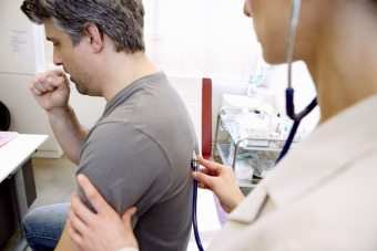
Frequent or worsening cough
We all cough from time to time, but if your cough becomes more frequent or chronic, it's time to see a doctor. COPD causes inflammation of the bronchioles and alveoli, making them less elastic. When this happens, the walls of the airways thicken and produce more mucus than normal. This leads to their blockage.
You will notice a mucous cough characteristic of pneumonia without other symptoms of pneumonia. If the mucus is no longer clear, this is a sign of a worsening condition.
Morning headaches
One of the symptoms of COPD is a dull, throbbing headache in the morning. The reason is that while you sleep, you don't breathe deeply enough, which leads to a buildup of carbon dioxide. At the same time blood vessels in the brain expand, causing headaches.
Many people do not associate headaches with COPD, but rather treat them as a separate symptom. But if you don't treat the underlying cause - the lack of oxygen entering the lungs during sleep - the headache will not go away.
Swollen ankles
 As COPD progresses, it leads to heart failure because the circulatory system does not receive enough oxygen. This can lead to fluid retention in the body, which is most easily identified in the ankles.
When the condition of the lungs deteriorates, the heart cannot push blood out with sufficient force. As a result, the blood supply to the kidneys and liver deteriorates, and they, in turn, cannot sufficiently perform the function of clearing toxins and getting rid of fluid. People suffer from similar swelling during flights and women during pregnancy.
Sleep problems
Do you prop yourself up with pillows to make your head and chest higher and make it easier for you to breathe? Do you sleep in a chair because it's easier for you to breathe in this position? Is it also possible that if you sleep on a flat surface, you experience dizziness upon waking up?
Since in horizontal position The lungs have a harder time working Many people with COPD do not sleep deeply, but they do not realize that this is due to problems with the lungs. They may also wake up with a cough.
If you regularly wake up with a cough or difficulty breathing, or feel weak, tired and possibly have a headache in the morning, talk to your doctor.
Barrel chest
One of the tests that doctors use to determine the development of COPD is to inhale while raising your arms above your head. Why? If this changes, known as an emphysematous (barrel-shaped) chest, then this is a concomitant symptom of COPD.
As a result of chronic inflammation, the lungs enlarge, pushing the diaphragm down. The chest wall also enlarges, weakening pectoral muscles and neck muscles and intercostal muscles.
When this happens, people with COPD involuntarily try to compensate by leaning forward while sitting, resting on their knees. This position stabilizes the chest and shoulders, making it easier to use the above muscles.
Bluish tint to lips and nails
Over time, if the blood does not carry enough oxygen throughout the body, lips and nails take on a bluish or grayish tint. Sometimes the bluishness is most pronounced in the sockets of the nails, and in some people the entire skin takes on a bluish or grayish tint.
The fact is that blood saturated with oxygen is bright red, and blood with less oxygen becomes darker and acquires a bluish tint. Your doctor will help you determine your blood oxygen levels.
Well, how not to miss pneumonia?
Be healthy!
One of the symptoms of COPD is a dull, throbbing headache in the morning. As COPD progresses, it leads to heart failure.
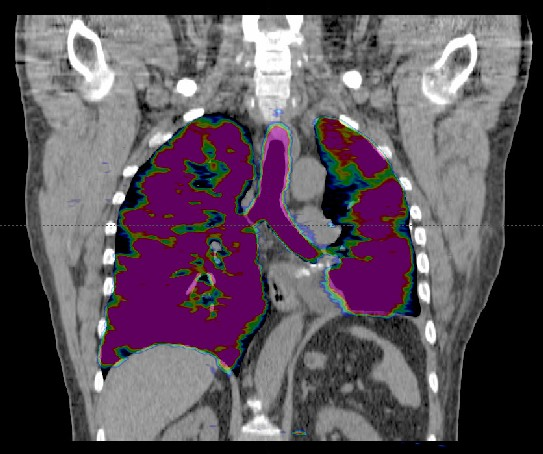
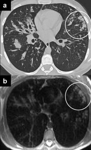
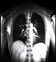
MRI of the lungs and bronchi is becoming an alternative method for diagnosing diseases respiratory system. Studying the structure of the lungs using MRI is based on receiving a response signal from protons in tissues and fluids human body, the so-called phenomenon of nuclear magnetic resonance.
Accurate and widely available in the future, this diagnostic method should be chosen when it is strictly necessary to avoid the use of ionizing radiation, in particular in children, pregnant women, as well as in diseases requiring repeated procedures (for example, neutropenia), which would contribute to a significant reduction in total radiation exposure doses.
What does it show
MRI has an advantage over CT in diagnosing the lungs in the following cases:
- Tumors (in the mediastinum and if they spread to the chest)
- Differential diagnosis atelectasis and tumors
- Differential diagnosis of mediastinal tumors
- Respiratory function assessment
- Diagnosis of pulmonary circulation disorders (embolism, hypoxic pulmonary vasoconstriction)
- Cystic fibrosis (with study of the vascular component)
MRI is as effective as CT in diagnosis for diagnoses:
- Pneumonia
- Atelectasis
- Pneumofibrosis (without study of the vascular component)
- Tuberculosis
- Pulmonary nodules (more than 3 mm)
- Sarcoidosis
- Acute and chronic pulmonary failure
- Pulmonary vascular abnormalities
- Pulmonary artery aneurysm
- Lung sequestration
- Arteriovenous malformations (Randu-Weber-Osler syndrome)
- Determining the stage of lung cancer
- Vasculitis (Wegener's disease)
- Pleural effusion of unknown origin
- Mesothelioma
In the following cases, it is better to diagnose the disease using CT, MRI is less effective:
- Pulmonary nodules (less than 3 mm)
- Interstitial lung disease
- Emphysema/COPD
Bronchial MRI is very effective for diagnosing bronchial wall thickening and dilatation of the central bronchi, but limitations in the resolution of the examination apparatus do not allow clear visualization of small vessels of the respiratory tract, such as the third or fourth generation.
The main type of diagnosis of bronchiectasis in adults, patients with cystic fibrosis and young patients is CT, but with repeated examinations, MRI may provide more complete information about the disease.
Although CT has better spatial resolution and shows morphology in more detail than MRI, bronchial MRI is better used to assess functional changes in hemodynamics and perfusion, as well as to assess ventilation and lung function.
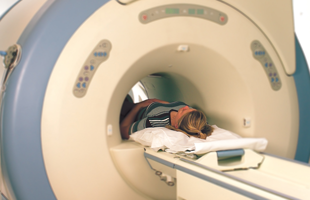
Preparation and contraindications
MRI of the lungs does not require special preparation. General requirements and contraindications can be found in the description of chest MRI.
MRI images of the lungs
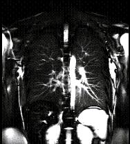
MRI images of the lungs in vascular mode
Price
The attending physician prescribes MRI for lung diseases if previous studies have not been able to provide complete data for making an accurate diagnosis. The research is quite expensive. The price of MRI of the lungs and bronchi varies from 3500 to 8000 rubles. It depends on whether an MRI is performed with or without contrast, the area of examination: lungs, bronchi, and also depends on the power of the device, the clinic and the region of residence.
Author:
Today we will tell you how to check your lungs. There are several ways. All of them will be discussed in detail in the article.
An organ of the body such as the lungs is located in chest. It is designed to support the breathing process. During it, air enters the body, which consists of many elements, one of them is oxygen. Oxygen saturation of the body is very important for the normal functioning of all systems.
Why check your lungs?
To assess a patient's health, a doctor needs to know how the lungs function regularly. Not only focal disorders can be determined using how the above-mentioned organ works, but also conclusions can be made regarding other disorders in the body.
For example, many heart diseases are directly related to impaired lung function. Therefore, as soon as the first signs of such problems appear, it may indicate a heart disease in a person.
Common diseases
Chronic lung diseases spread quickly. According to statistics, more than half of such diseases are observed. The most common painful deviations from the norm or pathology of the respiratory system:
- Pneumonia is an infectious disease of the lungs. It mainly affects the organs of people with weak immune systems.
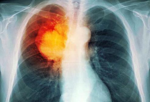
- Lung cancer or oncology is damage to organ cells by a malignant tumor.
- Due to an abnormal inflammatory response of lung tissue to external stimuli, the flow of air into the airways is limited, making it difficult for a person to breathe.
- Asthma. This is a chronic inflammatory disease of the respiratory tract.
- Bronchitis is a disease characterized by inflammation of the bronchi.
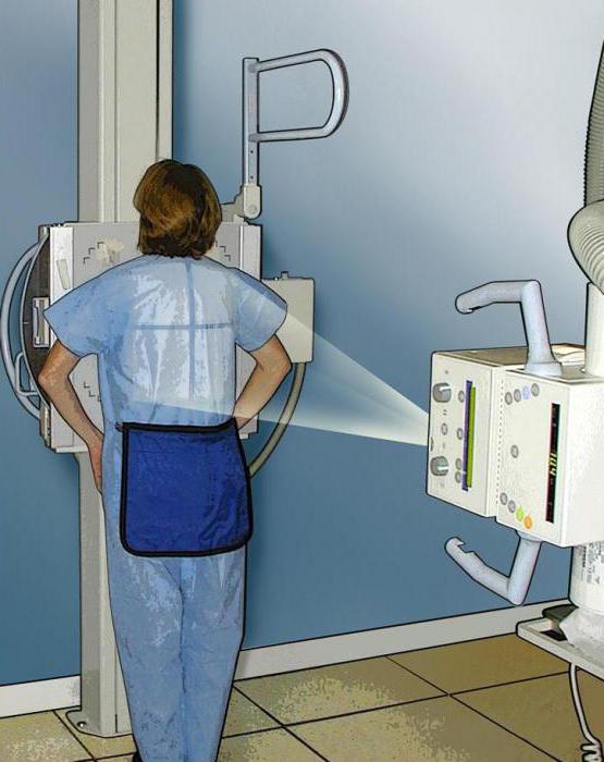
Inflammation and neoplasms in the lungs can appear not only in smokers or people living in areas with poor ecology. Therefore, organs need to be examined regularly, even if you feel well, an additional preventive measure will not harm. It is especially dangerous if the child is sick. How to check a child’s lungs for the presence of pulmonary diseases? You can start with a more common diagnosis. For example, this could be an x-ray of the lungs, the price this study not too high, about 200 rubles.
Today, various methods are used to diagnose the lungs and determine the disease and identify pathologies.
Types and methods of lung examination
The tidal volume of the lungs and the degree of respiratory failure can be determined using pulmonary ventilation.
Pleural puncture
The lining of the lungs is examined using a piece of tissue taken through a small puncture. Diagnosis is carried out under local anesthesia. If there is a suspicion of pleurisy, tumor or pleural effusions, then this procedure is prescribed.
Study of secretions
Mucous secretions from the respiratory tract are also examined to understand how properly the lungs are functioning. There are two methods: microscopic and bacterioscopic.
Conclusion
As you can see, an x-ray of the lungs, the price for which starts from 200 rubles, is not the only procedure that allows you to check the organ in detail. There are many more different methods.
If any alarming symptoms appear in the lung area, you should immediately consult a doctor. If this is the development of a pathology, then it needs to be treated in the early stages. Otherwise, it may affect the vital functions of other organs in the human body. Among other things, the earlier therapy is started, the lower the cost of medical services can be, since it is much easier to treat any pulmonary pathology at an early stage.
Often our illnesses make themselves felt by the appearance of previously absent symptoms. What should you do first if your lungs begin to act up? As a rule, everything starts with fluorography and sampling of mucus from the lungs.
Besides fluorography, how can you check your lungs?
The diagnostic process may involve a CT scan - computed tomography, which will show a cross-section of the lungs. The image will turn out colorless, but an experienced doctor will easily determine the presence of pathology, for example, oncology. The result can be expected half an hour after the procedure. The CT scan procedure is completely safe. During this process, a very small amount of radioactive rays is used, which cannot in any way affect the patient or staff. It is recommended not to eat food 4 hours before the test.
The spiral CT slice also has useful properties. The procedure involves moving a special apparatus around the patient's body. This machine scans internal organs and takes more than 100 frames. During such diagnostics, even the smallest tumor formations can be detected. The procedure lasts about a quarter of an hour.
The doctor may also need to examine the inside of the lungs. In this case, a biopsy is required. The latter is usually performed under local anesthesia, but general anesthesia can also be used. A flexible or rigid tube is used to collect material. A section obtained after a tomography will help determine whether a biopsy is needed.
Before the biopsy, you should not eat or drink water for several hours. The person is first given a sedative so that he can relax. It is also necessary to take a drug that reduces saliva production.
After the sedative has taken effect, an anesthetic is sprayed (in the case of local anesthesia). A bronchoscope is then inserted through the nose or mouth into the lungs. During the procedure, you can simultaneously observe organs and photograph pathologies, if any.
The procedure does not last long. After it, it is not recommended to eat food, at least until the effect of the anesthetic wears off. The person may be bothered for several days discomfort, but they go away on their own.
How can you check your lungs at home?
One of the common diseases is COPD - chronic obstructive pulmonary disease. This pathology is a combination of emphysema and obstructive bronchitis in a chronic form. It occurs due to smoking and damage to the respiratory system by irritating substances. COPD causes the lungs' ability to inhale oxygen to gradually become limited.
When this type of pathology develops in the lungs, symptoms appear slowly and are often difficult to recognize. COPD is a progressive pathology that cannot be left untreated. There are several signs that indicate that it is necessary to put the respiratory system in order.
How can you test a smoker's lungs?
- You need to take a deep breath and inflate the balloon with one exhalation. The resulting ball is the volume of our lungs. Normal - 3.5 liters;
- Blow out the candles. For this, cake candles are usually used. Their number should correspond to age. You need to blow them out one time from a distance of 70-80 cm;
- Hold your breath. Normally, you can hold your breath for at least a minute;
- Inflate a ball with a volume of 10 liters. This method is similar to the test with balloon. A woman’s full healthy breath is 2.5 liters. Accordingly, a woman whose lungs are normal can inflate a ball in 4 full deep breaths.
How to check lung capacity and function at home?
The first sign of pathology is shortness of breath. Many people ignore this symptom, attributing everything to age-related changes. However, the problem is that once COPD develops, it cannot be completely cured. This pathology can only be slowed down. If a person does not pay attention to the shortness of breath that occurs from normal walking, it means that he has less and less healthy lung tissue left. It is important to maintain and maintain lung function by increasing your activity level.
You can test yourself, for example, do some exercises, climb the stairs. It is worth experimenting with different types of activities and observing whether shortness of breath appears. Changes can be monitored over time. If it becomes increasingly difficult to take a full breath, you should consult a doctor.
How to check the bronchi and lungs yourself?
Frequent cough
All people cough periodically. However, frequent or chronic cough is a warning sign. COPD, for example, provokes inflammation of the alveoli and bronchioles, causing them to lose their elasticity. Subsequently, their walls thicken, more mucus is produced, and the lumens become clogged. With pathology, a cough with sputum production without other symptoms may occur. If the mucus has lost its transparency, it means that the person’s condition is worsening.
Morning headaches in smokers
An alarming sign is a throbbing headache that appears immediately after waking up and getting out of bed. It is due to the fact that a person breathes shallowly during sleep, as a result of which carbon dioxide accumulates, in addition, the blood vessels of the brain dilate.
Often headaches are not associated with pathologies of the respiratory system and are treated as a separate symptom. To get rid of it, it is necessary to eliminate the main cause - lack of oxygen.
Swelling of the ankles
When the bronchi are affected and there is no treatment, heart failure may develop because the circulatory system does not receive enough oxygen. As a result, fluid retention occurs in the body, which is primarily manifested by swelling in the feet and ankles.
As pulmonary pathology progresses, the heart pumps blood out with insufficient force, which affects the kidneys and liver. The latter, in turn, begin to poorly perform their functions of cleaning toxins and getting rid of fluid. Similar swelling is observed during pregnancy and during air travel.
Sleep problems
It is more difficult for the lungs to work in a horizontal position, so a sick person can place large pillows under his head and sleep reclining. After sleeping on a flat surface, you may feel dizzy. When lung health suffers, sick people cannot sleep deeply enough and often wake up coughing. If a person regularly wakes up for these reasons, feels tired, weak, or has a headache in the morning, then first of all one should suspect pathologies of the respiratory system.
Barrel chest
To determine COPD, doctors use one of the tests - inhaling while raising your arms above your head. In this position, in the presence of pathology, changes occur called emphysematous (barrel-shaped) chest.
Chronic inflammation causes the lungs to enlarge and the diaphragm to be pushed down. The chest wall enlarges, weakening the intercostal, pectoral and cervical muscles. Sick people involuntarily lean forward when sitting, leaning on their knees to compensate for the discomfort. This pose allows you to normalize the position of the shoulders and chest.
Cyanosis of lips, bluish tint of nails
With a lack of oxygen in the tissues, lips and nails acquire a grayish or bluish tint. Often, cyanosis is noticeable on the nail holes.

Sometimes absolutely the entire skin becomes grayish or bluish, which is quite noticeable even at home with the naked eye.
This is due to the fact that the blood, when sufficiently oxygenated, is bright red. When its deficiency occurs, it acquires a bluish tint and becomes darker.
A qualified specialist will definitely determine the level of oxygen in the blood during the diagnostic process.
Remember that if you suspect a serious pathology, you should immediately consult a doctor. The specialist will prescribe the necessary tests after the examination and interview.