It is possible to calculate the gestational age using ultrasound at an early stage with an accuracy of up to several days, which is especially important if the pregnant woman has made a mistake in the date of her period, and the amount of the hCG hormone does not coincide with the numbers prescribed for this period. It is necessary to determine the duration of pregnancy to timely determine various pathologies and the date of birth. How accurately an ultrasound determines the gestational age depends on when the woman came for her appointment. The sooner the better.
It is necessary to understand that even in the modern world of advanced technologies, a doctor can make a mistake in determining the duration of pregnancy, despite the ultra-modern equipment. One of the reasons for this is the individual characteristics of the body of both the mother and the fetus.
However, it is now more possible to accurately determine the date of birth than in the pre-technological era, when people made mistakes much more often, since the start of pregnancy was counted from the first day of the last menstrual period (gestational age or obstetric period). This is the reason why, according to most doctors, an official pregnancy lasts 40 weeks. But in reality it lasts 38 weeks, that is, 2 weeks shorter, since conception occurs in the middle of the cycle.
If we approach this issue historically, then obstetricians took into account the first day of menstruation primarily because the flow of menstrual blood is a rather dramatic process for a woman and it is much easier to remember or mark the start/end date on the calendar. In addition, there is a good chance that the doctor is not mistaken about the gestational age when the report starts from the date of the start of menstruation.
Another way to determine the day of pregnancy is when women calculate the time using a calculator. There are many calculators on the Internet that can help you determine the date of conception. To do this, you only need to enter a few values, the date of your last period and the length of your cycle. Of course, what such a calculator can show is significantly inferior to ultrasound in terms of calculation accuracy.
What does ultrasound determine?
Determining the gestational age using an ultrasound is much more accurate than using a calculator or period-based reading. It is known that people can make mistakes when relying on the values they put in the tables. The probability of error is especially high when there is a large amount of data.
But the errors are much smaller when a computer calculates. Modern ultrasound technology with the latest computer and software is able to take into account many parameters when determining the date of conception. In this case, the procedure for determining the date of conception and the date of probable birth is performed automatically, after which the ultrasound shows the ready-made values.
However, older generation ultrasound machines without modern software can be used to determine the date of conception. When performing an ultrasound, the doctor receives the following information:
- Number of fetuses and gestational sacs.
- Presence of fetal heartbeat.
- The dimensions of the fetus, from which the estimated gestational age is calculated.
- Abnormal features of the uterus, including abnormal shape and fibrous tissue.
Most ultrasounds are done transabdominally (through the skin of the abdomen) with a full bladder. If the pregnancy is up to six weeks, transvaginal ultrasound is used, which gives a better picture, since the fetus and fruit sac are not yet sufficiently developed at this time. During the examination, the bladder must be empty, and the method involves the use of a vaginal sensor.
How is the term calculated using ultrasound?
During the examination, you can see the following images on the ultrasound monitor, characteristic of a certain period:
- When an ultrasound scan is performed at 5 ½ weeks of gestation, a small fetal sac can be seen, but neither the embryo nor its heartbeat is visible. When the radiologist says 5 ½ weeks of gestation during an ultrasound examination, this is counted from the first day of the last menstrual period, which corresponds to 3 ½ weeks from conception.
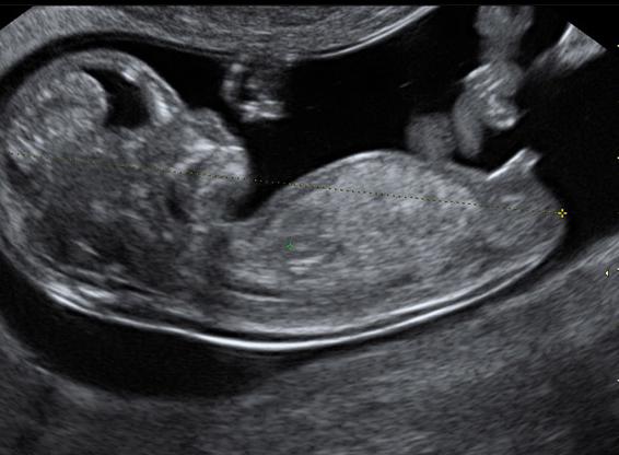
- From 6 to 7 weeks of gestation, the fetus is clearly visible on transvaginal ultrasound. In addition, his heartbeat is visible - from 90 to 110 beats/min. As the embryo gets older, the heart rate increases to 110-200 beats.
- At 8 weeks of gestation, the baby and its heartbeat are easily determined by both transvaginal and transabdominal ultrasound.
In the first trimester, an ultrasound scan first of all determines that the fetus is alive and establishes its age by measuring the length of the body from the crown to the beginning of the spine. Such measurements are a reliable indicator of the gestational age of the fetus. Therefore, this is the main method of how ultrasound determines the gestational age.

There are many tables for calculating age depending on this length. But there is a simple rule that from 6 to 11 weeks the embryo grows at a rate of 1 mm per day. From this, the gestational age of the embryo can be calculated. It is equal to 6 weeks + the number of millimeters from the crown to the coccyx, expressing days.
For example, if the embryo measures 23 mm between 6 and 11 weeks, its age is:
6 weeks + 23 mm (or 23 days) = 6 weeks. + 3 weeks +2 days = 9 weeks and 2 days
Based on these values, the date of birth is calculated using ultrasound. The accuracy of calculations of what period is indicated by ultrasound decreases after 12 weeks and is replaced by calculations using measurements of the size of the embryo's head (biparietal diameter). But it should be expected that with such a duration of pregnancy, an error is possible: according to ultrasound, the period may be longer or shorter.
Should you trust ultrasound?
How accurately an ultrasound determines the gestational age depends on what time the diagnosis is made. An ultrasound to determine the due date, which was performed at 12 weeks of pregnancy, usually has an error of 3-5 days. The most accurate is ultrasound from 8 to 11 weeks of gestation, since at this time the fetus develops very quickly and changes in its size are visible, which are used to determine the period. In addition, at this time the fetal body is easier to measure because it does not bend or twist in the same way as is typical at a later age.

Ultrasound after 22 weeks of gestation cannot be used at all to calculate the due date, since during this period the size of the baby and its age are no longer related. The magnitude of the error with the gestational age at this stage is very large and amounts to 2-3 weeks. In this case, the term according to ultrasound is often set to be greater than the obstetric term, or the term according to ultrasound is less than the obstetric one.
Thus, if a pregnant woman wants to use ultrasound to calculate her due date, the most accurate days will be around the 8th week of pregnancy. In this case, it is advisable to use the latest equipment: with outdated equipment, only an experienced sonologist can accurately determine the gestational age using ultrasound. At the same time, when examining using new equipment, much depends on the quality of computer software and equipment. Therefore, the point is not whether ultrasound can be wrong, but by how much and why.
Search for pathologies
In addition to determining the timing of pregnancy by ultrasound, during the examination the doctor needs to check for certain abnormalities in the baby. Diagnosis for the presence of abnormalities is carried out between 10 and 14 weeks of pregnancy. This study is performed using transabdominal ultrasound. If the picture is unclear, a transvaginal examination may be necessary.
During an ultrasound, a complete assessment of the pregnancy is carried out, for which the amount of amniotic fluid, the position and type of the placenta, and a detailed image of the embryo are determined. The fruit is measured in all respects. The presence of chromosomal abnormalities is indicated by a rounder head shape, a short nasal bone, wide hands with short fingers and other signs.
Thus, an ultrasound aimed at searching for abnormalities is a very important examination. This is why doctors recommend that every pregnant woman have an ultrasound at 12 weeks. Even the first scan may show chromosomal abnormalities. For example, thickening of the collar zone (in the neck area), which makes one suspect Down syndrome and other genetic problems.

The transparency of the fetal nuchal zone is the amount of fluid that has accumulated under the skin in the back of the fetal neck. The word "transparency" is used here because in the image it appears as a black area just under the skin at the back of the neck. The amount of this fluid may increase with Down syndrome and other genetic abnormalities. Currently, ultrasound is the most reliable non-invasive method for determining Down syndrome in an unborn child between 11 and 14 weeks of pregnancy.
Echogenic sites in the heart also increase the risk of Down syndrome. To clarify the diagnosis, if the collar area is transparent, the presence of a pronounced nasal bone is checked. In 75% of fetuses with Down syndrome it is not clearly expressed. Therefore, a visible nasal bone reduces the likelihood of this pathology.
During the ultrasound test of the nuchal area, it is also checked whether the child has all the limbs, whether the head and brain are normally developed, whether the stomach and bladder are clearly visible, and whether the umbilical cord is positioned correctly. From the 12th week, ultrasound allows you to clearly view the spine to exclude an anomaly such as spina bifida.
If the fetal intestine appears bright and white (the same color as the bones), it is called echogenic intestine. Its appearance can be caused by various reasons, and increases the risk of a congenital disease such as chromosomal trisomy. The same pathology is indicated by the short femur and humerus, as well as the choroid plexus cyst, which is located in the fetal brain and disappears by the 28th week of pregnancy. If present, it increases the risk of chromosomal trisomy.
Ultrasound allows one to identify pathologies such as dilated renal hilum (pyelectasis), which can subsequently lead to pyelonephritis. However, you shouldn’t despair right away: when asked whether an ultrasound can be wrong, many women who have given birth to a healthy child will give an affirmative answer. However, to confirm the diagnosis, it will be necessary to undergo additional examinations, after which the doctor will make a verdict.
Pregnancy is one of the most beautiful periods in the life of a representative of the fair sex. It is worth noting that medicine knows two options for calculating the time of gestation in the uterus: the obstetric gestational age and the real one.
Where does it all begin?
To begin with, it is worth talking about how fertilization occurs. Around the middle of the month, the female egg leaves the follicle and slowly moves along. This is where it meets the male cell. The chromosomes then fuse and conception occurs. Having descended into the uterine muscle, the fertilized egg penetrates the endometrium, and from this moment we can assume that the pregnancy has taken place.
Determination of gestational age
When a woman realizes that she is in an interesting position, her initial task is to determine the timing. The gestational age is calculated by week. Typically, the period of time during which the baby is in the mother's womb is 40 weeks. A slight shift in one direction or another is considered normal and does not require any correction. Doctors distinguish between the obstetric gestational age and the real one.

Real gestation time
This period starts from the moment when ovulation occurred. The release of the egg from the follicle is the day from which the actual gestational age is calculated. Most women's clinics that monitor the course of pregnancy use this method of calculation. If you decide to take a blood test to determine the content in it, you will also be provided with a result that indicates the real value of the period.
Obstetric gestational age
This time period begins its countdown from the first day of the last bleeding from the woman’s genital tract. This date is used to calculate the expected date of birth of the baby. Also, many representatives of the fair sex use this method to determine the duration of pregnancy. This is why women so often have discrepancies with the calculation made by the doctor.
Obstetric gestational age and real
In most cases, the difference between these counting methods is two weeks. In a standard female cycle of twenty-eight days, the release of the egg from the ovary occurs exactly two weeks after the start of the last menstruation.
However, not all representatives of the fair sex have a standard cycle length. For example, some women ovulate one week after the start of their last period. In such cases, the difference between the obstetric and actual terms will be one week.
If a woman’s egg was released three weeks after the start of her last menstruation, then in this case the obstetric pregnancy period and the real one will have a difference of twenty-one days.
All situations described are normal. That is why the gestational age by week should be set taking into account the length of the woman’s menstrual cycle. It is impossible to equate all representatives of the fair sex to generally accepted standards. This can lead to incorrect calculation of the baby's gestation period.
pregnancy by ultrasound

There are situations when a woman cannot name the date of the first day of her last menstruation. This situation often occurs if a woman has recently given birth or is breastfeeding. In such cases, representatives of the fair sex are recommended to undergo diagnostics using an ultrasound machine (ultrasound).
A short period of pregnancy, which is not yet possible to determine by manual examination, is easily diagnosed by ultrasound. It is worth noting that a specialist can determine the presence of a woman in the uterus starting from the fourth obstetric week. All measurements and determinations of the period are calculated using the obstetric method.

Instead of a conclusion
If you don't know how to calculate your gestational age, ask your doctor. In most cases, it is enough to know the date of the last menstruation and the length of the female menstrual cycle. If necessary, an ultrasound examination is also prescribed. Carry out the calculation using the same method used by the specialist. Only in this case you will not encounter discrepancies and will not find yourself in a controversial situation.
Gestational age- one of the main components that a gynecologist needs to know about for proper management of pregnancy. After all, on the basis of this, the doctor will prescribe the necessary examinations, tests, routine ultrasounds, monitor intrauterine growth and development of the fetus, and identify possible violations during the course of the pregnancy. In addition, determining the due date allows you to fairly accurately calculate the date of birth and the date of maternity leave. It is necessary for the woman herself to know the duration of her pregnancy in order to mentally prepare for childbirth, have time to prepare a room or corner for the unborn baby, and purchase all the necessary accessories and clothes for him.
In this article we will look at how to determine the duration of pregnancy.
Calculate by monthly
For a woman who comes to register at the antenatal clinic, the gynecologist first of all asks a question about when her last period began. It is from this date that he begins to keep a record, despite the fact that fertilization has not yet occurred. It is known that fertilization of the egg occurs on the day of ovulation, which occurs in the middle of the menstrual cycle. This method of determining the gestational age is called “obstetric”.
An online calculator will help determine the period:
How to determine the gestational age by the date of conception
It is known that conception occurs only during ovulation within 24 hours after the egg leaves the follicle. And the viability of sperm in the female genital tract is up to 3 days. It turns out that the date of sexual intercourse does not always coincide with the date of conception.
Ovulation occurs in the middle of the menstrual cycle. A woman who monitors her menstrual cycle can quite accurately calculate the onset of ovulation. In addition, many at this time develop characteristic symptoms: aching pain in the lower abdomen, breast swelling, irritability, copious vaginal discharge, increased libido.
It is worth noting that only those women who have a regular menstrual cycle can determine the gestational age by the date of conception.
Online calculator:
(calculation will take a few seconds)
How to calculate using ultrasound
 It is possible to find out the gestational age using the results of an ultrasound examination (ultrasound). It is determined according to the size of the fetus. The first ultrasound of the expectant mother is performed at 12-14 weeks, but it is still impossible to determine the exact date, since the development of the fetus occurs individually in each woman. It is possible to find out the age of the fetus by ultrasound with an accuracy of one day only in the first weeks after conception.
Determination during examination by a gynecologist
A gynecologist can determine a woman's gestational age by the size of her uterus during a gynecological examination. At 5-6 weeks, the size of the uterus can be compared to a chicken egg, at 8 weeks - to a goose egg. Later, it will be more difficult to determine how many weeks a woman has based on the size of her uterus.
Find out the due date by the first fetal movement
In the first pregnancy, a woman begins to feel fetal movements at 20 weeks, in the second - at 18 weeks. But this method does not always allow one to determine the period, since some expectant mothers may feel the first movements of the fetus earlier or later than the indicated weeks - some, due to the structural features of the body, cannot feel the movements of the fetus, others confuse the work of the intestines (increased gas formation) with movements.
Determine by hCG
Human chorionic gonadotropin (hCG)- one of the most important indicators indicating the onset of pregnancy and its normal development. The hormone begins to be produced in the female body after implantation of the embryo into the wall of the uterus. The level of hCG in the blood allows you to find out the approximate age of the fetus. With multiple pregnancy, the level of the hormone in the blood increases in proportion to the number of fetuses.
| Week of pregnancy | HCG level, honey/ml |
| 1 - 2 | 25 - 156 |
| 2 - 3 | 101 - 4870 |
| 3 - 4 | 1110 - 31500 |
| 4 - 5 | 2560 - 82300 |
| 5 - 6 | 23100 - 151000 |
| 6 - 7 | 27300 - 233000 |
| 7 - 11 | 20900 - 291000 |
| 11 - 16 | 6140 - 103000 |
| 16 - 24 | 4720 - 80100 |
| 21 - 39 | 2700 - 78100 |
A woman may have suspicions about a possible “interesting situation” long before her next menstruation is missed. Modern test strips can determine the content of the specific hormone hCG in the urine already on the first day of the delay, and some even several days before it. Whatever the test result, a woman wants to make sure she is pregnant as early as possible. This article will tell you when the baby can be seen for the first time on an ultrasound.

Minimum terms for determination
After conception has taken place, intensive processes begin inside the expectant mother, which she most often is not aware of. On the very first day, the fertilized egg divides and moves through the fallopian tube, where conception took place, into the uterine cavity. This journey lasts about four days. It is no longer a set of individual cells that descends into the uterus, but a blastocyte - a ball-shaped formation. It penetrates the lining of the uterus. This is implantation. This happens 6-7 days after fertilization, and sometimes a woman feels implantation by slight pulling sensations in the lower abdomen.

The earliest symptom of pregnancy is sometimes the so-called implantation bleeding - a few drops of bloody or bloody discharge at the time of blastocyte implantation into the endometrium. This doesn't mean it's time to run out for a test or sign up for an ultrasound.
The test strips react to the formation of the so-called pregnancy hormone - hCG, but it is just beginning, the level of the hormone is below the control level of the sensitivity of the test strips. But a blastocyte cannot be seen on an ultrasound - its size is only 0.2 mm.

How is an ultrasound done?
To determine pregnancy, two types of ultrasound examination are used - transvaginal and transabdominal. In the first case, the doctor examines the uterine cavity and its contents with a vaginal sensor. In the second case, the inspection is carried out with a sensor through the abdominal wall. For the most part, doctors prefer the first method when it comes to early pregnancy. Through the vagina it is much easier to see the embryo and its structure.
An abdominal ultrasound of the pelvic organs is recommended to be performed with a full bladder, a transvaginal ultrasound with an empty one, and it is better to take care in advance that the intestines are not distended from gases. To do this, a few hours before going to the doctor, it is advisable for a woman to take Espumisan or Smecta.


It should be noted that using the transvaginal method, pregnancy can be seen earlier than the transabdominal method, by several days. Thus, a vaginal sensor and a good specialist in addition can tell a woman about her “interesting situation” already on the 5-6th day from the day of the delay, and a scan through the abdomen may not show pregnancy even on the 8-10th day. The procedure is painless, non-hazardous for the woman and baby, and lasts no more than 5-7 minutes.

Transcript of the first ultrasound
At the very first ultrasound examination to determine pregnancy, the diagnostician will be able to detect an echogenic formation. This is the fertilized egg. Its size will indicate the exact stage of pregnancy. The doctor will also determine the size of the yolk sac, the position of the fertilized egg, the thickness of the endometrium, and rule out inflammatory processes in it, as well as the presence of cysts, polyps and other unwanted formations. The dimensions of the fertilized egg and the timing table are presented below.

Are errors possible?
The ultrasound diagnostic method is considered one of the most accurate for determining pregnancy in the early stages, but you should not assume that its accuracy is 100%. In gynecology, the accuracy of this test is estimated at approximately 90%. In early pregnancy, accuracy decreases to 75%. A doctor is, first of all, a person, and not a machine with a program embedded in it. He has the right to make mistakes, especially if a woman has problems with the health of her reproductive system. Thus, a doctor may confuse uterine fibroids with pregnancy in the early stages if the woman had not previously been diagnosed with fibroids and only learned about its presence through an ultrasound. A cyst or polyp can be confused with a fertilized egg, since a cyst is also an echogenic formation.

If a woman had late ovulation, then the pregnancy a week after the delay may not be detected at all by an ultrasound diagnostic specialist, since the fertilized egg later descended into the uterus and is not yet visualized. Naturally, the doctor will write in the conclusion that no signs of pregnancy were found, but after 7-10 days, during a repeat examination, he will be able to determine both the fertilized egg and its structure. Only the size will help you understand that ovulation was indeed late.

Common Questions
On the Internet, inexperienced pregnant women and those who are still dreaming of an “interesting situation” ask many questions regarding the earliest diagnosis. It is worth talking about the most common situations in more detail.
The pregnancy test was positive, but the ultrasound was not.
There may be several reasons for this. First of all, one should not rule out that the test turned out to be defective; this happens, and quite often, especially when it comes to inexpensive test strips, which are sold on almost every corner. In their desire to see the two treasured stripes, some ladies go too far, starting to look for “ghost” stripes on the dough strips. If they find it, they automatically begin to consider their test positive, although in reality there may not be a pregnancy.
If the test still did not deceive, then the reason for the negative conclusion of the ultrasound diagnostic doctor may be that the woman went to the doctor too early, and the fertilized egg is not yet visible. The device itself may be outdated, with low sensitivity and poor resolution. The reason for the absence of signs of pregnancy on ultrasound may be late ovulation, the presence of an inflammatory process in the uterine cavity, and, of course, insufficient qualifications of the doctor.

The pregnancy test was negative, but the ultrasound was positive
There may also be plenty of reasons for this situation. Firstly, the woman could have carried out the test at home with an error, the test could have been defective or expired, and it is also possible that it was carried out too early, when the level of the hCG hormone in the urine was still insufficient for the test to respond brightly. second stripe.
Ultrasound diagnosis in this case is rarely premature, since a woman, after a negative home test, does not rush to the doctor, patiently waiting for the onset of a late period. After one and a half to two weeks of delay When the lady finally goes to the doctor, the pregnancy is already clearly visible on an ultrasound. Therefore, ultrasound results should be considered more reliable than home test results. In doubtful cases, you can donate blood for hCG to obtain even more accurate data.

How to calculate gestational age using ultrasound?
To do this, you can use the table above. If greater detail of the period is required, use a table of correspondence of the period, accurate to the day, to the average internal diameter of the fertilized egg (SVD). A table of pregnancy periods in accordance with the SVD is given below.
The value of the average internal diameter of the ovum | Gestational age |