Temporal bone (os temporale) steam room, part of the base and side wall of the skull between sphenoid bone anteriorly and the occipital bone posteriorly. It houses the organs of hearing and balance. The temporal bone is divided into pyramid, tympanic and squamosal parts.
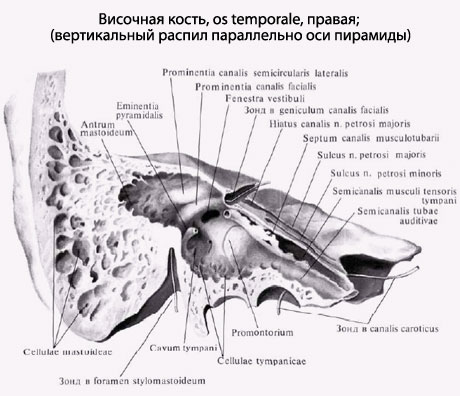
The pyramid, or rocky part (pars petrosa), has a triangular shape and is located obliquely in the horizontal plane. The apex of the pyramid is directed forward and medially, and the base is directed backward and laterally. At the top of the pyramid is the internal opening of the carotid canal (canalis caroticus). Nearby and to the lateral side is the muscular-tubal canal (canalis musculotubarius), which is divided by a septum into two semi-canals: the semi-canal of the auditory tube (semicanalis tubae auditivae) and the semi-canal of the tensor tympani muscle (semicanalis musculi tensoris tympani).
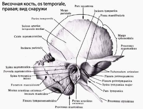
The pyramid has three surfaces: front, back and bottom. Front surface The pyramid faces up and forward. Near the apex on this surface there is a small trigeminal impression (impressio trigemini). Two openings are visible lateral to this depression. The larger of them is called the cleft (hole) of the canal of the greater petrosal nerve (hiatus canalis nervi petrosi majoris), from which a narrow groove of the same name runs forward and medially. Anterior and lateral is the cleft of the lesser petrosal nerve (hiatus canalis nervi petrosi minoris), which passes into the groove of this nerve. On the front surface of the pyramid there is a flattened section - the roof of the tympanic cavity (tegmen thympani), which is its upper wall. Along the upper edge of the pyramid there is a groove of the superior petrosal sinus (sulcus sinus petrosi superioris).
![]()
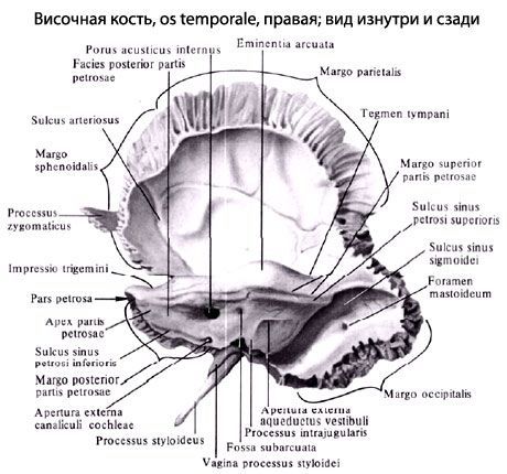
Back surface of the pyramid facing posteriorly and medially. In the middle of this surface is the internal auditory opening (porus acusticus internus). It leads into the internal auditory canal (medtus acusticus internus). Lateral and slightly above this opening is the subarc fossa (fossa subarcuata), below and lateral to which there is a little noticeable external aperture (hole) of the vestibular aqueduct (apertura externa aqueductus vestibuli). A groove of the inferior petrosal sinus (sulcus sinus petrosi inferioris) runs along the posterior edge of the pyramid. At the lateral end of this groove, adjacent to the jugular fossa, there is a depression, at the bottom of which the external aperture of the cochlear canaliculus (apertura externa canaliculi cochleae) opens.
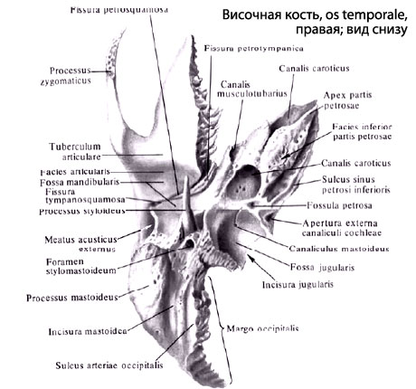
Bottom surface of the pyramid has a complex terrain. Near the base of the pyramid there is a deep jugular fossa (fossa jugularis). Anterior to it there is a rounded external opening of the carotid canal, inside of which, in its wall, there are 2-3 openings of the carotid-tympanic tubules connecting the carotid canal with the tympanic cavity. On the ridge between the jugular fossa and the external opening of the carotid canal there is a small lobe (fossula petrosa). Lateral to the jugular fossa, a thin and long styloid process (processus styloideus) is directed downwards. Behind the process there is a stylomastoid foramen (foramen stylomastoideum), and behind this opening a wide, easily palpable mastoid process (processus mastoideus) is directed downwards.

In the thickness of the mastoid process there are air-filled cells. The largest cell, the mastoid cave (Antrum mastoideum), communicates with the tympanic cavity. Medially, the mastoid process is limited by the deep mastoid notch (incisure mastoidea). Medial to this notch is the groove of the occipital artery (sulcus arteriae occipitalis). At the base of the mastoid process there is sometimes a mastoid foramen (foramen mastoideum).
The tympanic part (pars tympanica) is formed by a curved narrow bone plate, which in front, below and behind limits the external auditory opening (porus acusticus externus), leading into the external auditory canal (meatus acusticus externus). Between the tympanic part and the mastoid process there is a narrow tympanomastoid fissure (fissure tympanomastoidea). In front of the external auditory opening is the tympanic-squamous fissure (fissure tympanosquamosa). A narrow bone plate protrudes into this gap from the inside - the edge of the roof of the tympanic cavity. As a result, the tympanic-squamous fissure is divided into the anterior stony-squamous fissure (fissura petrosquamosa) and the petrotympanic fissure (fissura petrotympanica, Glaser's fissure), through which a branch of the facial nerve, the chorda tympani, emerges from the tympanic cavity.
The scaly part (pars squamosa) is a convex plate outward, having a beveled free upper edge for connection with the parietal bone and the greater wing sphenoid bone. The outer temporal surface of the scales is smooth. On the inner cerebral surface of the scales there are cerebral elevations, finger-like impressions and arterial grooves. From the scales, above and anterior to the external auditory canal, the zygomatic process (processus zygomaticus) begins. Connecting with the temporal process of the zygomatic bone, it forms the zygomatic arch. Behind the zygomatic process, at its base, is the mandibular fossa (fossa mandibularis) for articulation with the condylar process of the mandible to form the temporomandibular joint.
Canals of the temporal bone. Several channels of the temporal bone pass through the pyramid for cranial nerves and blood vessels.
The carotid canal canalis cardticus) begins on the lower surface of the pyramid with the external carotid foramen, goes upward, bends almost at a right angle, then goes medially and forward. The canal ends with the internal carotid foramen at the top of the pyramid of the temporal bone. The internal carotid artery and the nerves of the carotid plexus pass through this canal into the cranial cavity.

Carotid-tympanic tubules (canaliculi caroticotympanic!), numbering 2-3, depart from the carotid canal and are directed into the tympanic cavity. These tubules contain arteries and nerves of the same name.
The muscular-tubal canal (canalis musculotubarius) begins at the top of the pyramid of the temporal bone, goes back and laterally and opens into the tympanic cavity. A horizontal partition divides it into two parts. Above is the hemicanal of the tensor tympani muscle (semicanalis musculi tensoris tympani), containing the muscle of the same name. Below is the semicanal of the auditory tube (semicanalis tubae auditivae).

The facial canal (canalis facialis) begins in the internal auditory canal. It first runs transverse to the long axis of the pyramid to the level of the cleft of the greater petrosal nerve canal. Having reached the cleft, the canal forms a knee, then is directed at a right angle back and laterally. Having passed along the medial wall of the tympanic cavity, the canal turns vertically downwards and ends with the stylomastoid foramen. The facial nerve passes through this canal.
The canaliculus chordae tympani comes from the wall of the facial canal in its final section and opens into the tympanic cavity. The nerve, the chorda tympani, passes through this canal.
The tympanic canaliculus (canaliculus tympanicus) begins at the bottom of the stony fossa, goes upward, and pierces the wall of the tympanic cavity. The canaliculus then passes along its medial wall and ends in the area of the cleft of the canal of the lesser petrosal nerve. The tympanic nerve passes through this canaliculus.
The mastoid tubule (canaliculus mastoideus) begins in the jugular fossa and ends in the tympanomastoid fissure. The auricular branch of the vagus nerve passes through this canaliculus.
Temporal bone, os temporale, a paired bone, has a complex structure, as it performs all 3 functions of the skeleton and not only forms part of the side wall and base of the skull, but also contains the organs of hearing and gravity. It is the product of the fusion of several bones (mixed bone), which independently exist in some animals, and therefore consists of three parts:
- scaly part, pars squamosa;
- drum part, pars tympanica and
- rocky part, pars petrosa.
During the 1st year of life, they merge into a single bone, closing the external auditory canal, meatus acusticus externus, in such a way that the scaly part lies above it, the stony part medially from it, and the tympanic part behind, below and in front. Traces of fusion individual parts of the temporal bone are preserved for life in the form of intermediate sutures and fissures, namely: at the border of pars squamosa and pars petrosa, on the anterosuperior surface of the latter - fissura petrosquamosa; in the depths of the mandibular fossa - fissura tympanosquamosa, which is divided by a process of the petrous part into fissura petrosquamosa and fissura petrotympanica (the chorda tympani nerve exits through it).

Scaly part, pars squamosa, participates in the formation of the lateral walls of the skull. It belongs to the integumentary bones, i.e. it ossifies on the basis of connective tissue and has a relatively simple structure in the form of a vertical plate with a rounded edge overlapping the corresponding edge of the parietal bone, margo squamosa, in the form of fish scales, which is where its name comes from.
On its cerebral surface, facies cerebralis, visible traces of the brain, finger impressions, impressiones digitatae, and an upward groove from a. meningea media. The outer surface of the scales is smooth, participates in the formation of the temporal fossa and is therefore called facies temporalis. The zygomatic process, processus zygomaticus, departs from it, which goes forward to connect with the zygomatic bone. At its origin, the zygomatic process has two roots: anterior and posterior, between which there is a fossa for articulation with the lower jaw, fossa mandibularis.

An articular tubercle, tuberculum articulare, is placed on the lower surface of the anterior root, which prevents the head of the lower jaw from dislocating forward when the mouth opens significantly.

Part of the tympanum, pars tympanica, temporal bone forms the anterior, lower and part of the posterior edge of the external auditory canal, ossifies endesmally and, like all integumentary bones, has the appearance of a plate, only sharply curved. The external auditory canal, meatus acusticus externus, is a short canal that goes inward and somewhat forward and leads into the tympanic cavity. The upper edge of its external opening, poms acusticus externus, and part of the posterior edge are formed by the scales of the temporal bone, and along the remaining length by the tympanic part.
In a newborn, the external auditory canal is not yet formed, since the tympanic part is an incomplete ring (annulus tympanicus), covered by the eardrum. Due to such a close position of the eardrum outward in newborns and children early age Diseases of the tympanic cavity are more common. The petrous part, pars petrosa, is so named for the strength of its bone substance, due to the fact that this part of the bone is involved in the base of the skull, and is the bony seat of the organs of hearing and gravity, which have a very delicate structure and require strong protection from damage. It develops on the basis of cartilage. The second name of this part is the pyramid, given by its shape as a triangular pyramid, the base of which faces outward, and the apex faces forward and inward towards the sphenoid bone.
The pyramid has three surfaces: front, back and bottom. The anterior surface is part of the bottom of the middle cranial fossa; the posterior surface faces posteriorly and medially and forms part of the anterior wall of the posterior cranial fossa; the lower surface faces down and is visible only on the outer surface of the base of the skull. The external relief of the pyramid is complex and is determined by its structure as a container for the middle (tympanic cavity) and inner ear (bone labyrinth, consisting of the cochlea and semicircular canals), as well as the passage of nerves and blood vessels. On the anterior surface of the pyramid, near its apex, there is a noticeable small depression, impressio trigemini, from the trigeminal ganglion (n. trigemini). Two thin grooves run outward from it, the medial one is sulcus n. petrbsi majoris, and lateral - sulcus n. petrosi minoris. They lead to two foramina of the same name: the medial one, hiatus canalis n. petrosi majoris, and lateral, hiatus canalis n. petrosi minoris. Outside of these openings, an arched elevation, eminentia arcuata, is noticeable, formed due to the protrusion of the rapidly developing labyrinth, in particular the superior semicircular canal.
The surface of the bone between eminentia arcuata and squama temporalis forms the roof of the tympanic cavity, tegmen tympani. Approximately in the middle of the posterior surface of the pyramid there is an internal auditory opening, porus acusticus internus, which leads to the internal auditory canal, meatus acusticus internus, where the facial and auditory nerves, as well as the artery and veins of the labyrinth, pass. From the lower surface of the pyramid, facing the base of the skull, extends a thin pointed styloid process, processus styloideus, which serves as the attachment point for the muscles of the “anatomical bouquet” (mm. styloglossus, stylohyoideus, stylopharyngeus), as well as ligaments - ligg. stylohyoideum and stylomandibular. The styloid process represents a part of the temporal bone of branchial origin. Together with lig. stylohyoideum it is a remnant of the hyoid arch. Between the styloid and mastoid processes there is a stylomastoid foramen, foramen stylomastoideum, through which n. facialis and a small artery enters. Medial to the styloid process there is a deep jugular fossa, fossa jugularis. Anterior to the fossa jugulalis, separated from it by a sharp ridge, is the external opening of the carotid canal, foramen caroticum externum.

The pyramid has three edges: anterior, posterior and upper. The short anterior margin forms an acute angle with the scales. In this corner, the opening of the myotubal canal, canalis musculotubarius, leading into the tympanic cavity is noticeable. This channel is divided by a partition into two sections: upper and lower. Upper, smaller, semicanal, semicanalis m. tensoris tympani, houses this muscle, and the lower, larger one, semicanalis tubae auditivae, is the bony part of the auditory tube, which serves to conduct air from the pharynx into the tympanic cavity. Along the upper edge of the pyramid, separating the anterior and posterior surfaces, there is a clearly visible groove, sulcus sinus petrosi superiors, a trace of the venous sinus of the same name. The posterior edge of the pyramid anterior to the fossa jugularis connects with the basilar part of the occipital bone and, together with this bone, forms the sulcus sinus petrosi inferioris - a trace of the inferior petrosal venous sinus.

The outer surface of the base of the pyramid serves as a place for muscle attachment, which determines its external relief (process, notches, roughness). Downwards it extends into the mastoid process, processus mastoideus. The sternocleidomastoid muscle is attached to it, which maintains the head in the balance necessary for an upright position of the body. Therefore, the mastoid process is absent in quadrupeds and even apes and develops only in humans in connection with their upright posture. On the medial side of the mastoid process there is a deep mastoid notch, incisura mastoidea, - the place of attachment of m. digastricus; even more inward - a small groove, sulcus a. occipitalis, - trace of the artery of the same name. On the outer surface of the base of the mastoid process, a smooth triangle is distinguished, which is a place for rapid access to the cells of the mastoid process when they are filled with pus.
Inside the mastoid process contains these cells cellulae mastoideae, which are air cavities separated by bone bars that receive air from the tympanic cavity, with which they communicate through the antrum mastoideum. On the cerebral surface of the base of the pyramid there is a deep groove, sulcus sinus sigmoidei, where the venous sinus of the same name lies. Canals of the temporal bone. The largest canal is the canalis caroticus, through which the internal carotid artery passes. Beginning with its external opening on the lower surface of the pyramid, it rises upward, then bends at a right angle and opens with its internal opening at the apex of the pyramid medial to the canalis musculotubarius.
The facial canal, canalis facialis, begins in the depths of the porus acusticus internus, from where the canal first goes forward and laterally to the cracks (hiatus) on the anterior surface of the pyramid; at these holes, the canal, remaining horizontal, turns at a right angle laterally and backward, forming a bend - the geniculum canalis facialis, and then downwards and ends through the foramen stylomastoideum, located on the lower surface of the pyramid of the temporal bone.
Which doctors should I contact to examine the temporal bone:
Traumatologist
What diseases are associated with the temporal bone:
What tests and diagnostics need to be done for the Temporal bone:
CT scan of the skull
MRI of the skull
X-ray of the skull
Is something bothering you? Do you want to know more detailed information about the Temporal bone or do you need an examination? You can make an appointment with a doctor– clinic Eurolab always at your service! The best doctors will examine you, advise you, provide necessary help and make a diagnosis. You can also call a doctor at home. Clinic Eurolab open for you around the clock.
How to contact the clinic:
Phone number of our clinic in Kyiv: (+38 044) 206-20-00 (multi-channel). The clinic secretary will select a convenient day and time for you to visit the doctor. Our coordinates and directions are indicated. Look in more detail about all the clinic’s services on it.
If you have previously performed any research, Be sure to take their results to a doctor for consultation. If the studies have not been performed, we will do everything necessary in our clinic or with our colleagues in other clinics.
It is necessary to take a very careful approach to your overall health. There are many diseases that at first do not manifest themselves in our body, but in the end it turns out that, unfortunately, it is too late to treat them. To do this, you just need to do it several times a year. be examined by a doctor, in order not only to prevent a terrible disease, but also to maintain a healthy spirit in the body and the organism as a whole.
If you want to ask a doctor a question, use the online consultation section, perhaps you will find answers to your questions there and read self care tips. If you are interested in reviews about clinics and doctors, try to find the information you need on. Also register on the medical portal Eurolab to stay up to date latest news and updates to information about the Temporal Bone on the website, which will be automatically sent to you by email.
Other anatomical terms starting with the letter "B":
| Upper esophageal sphincter |
| Laryngeal prominence |
| Vagina |
| Hair |