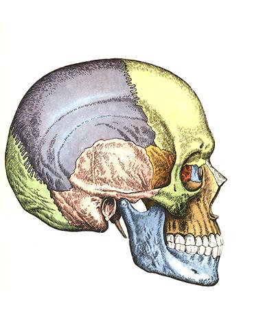
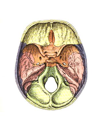
TEMPORAL BONE
Temporal bone, os tempordle, a paired bone, has a complex structure, as it performs all 3 functions of the skeleton and not only forms part of the side wall and base of the skull, but also contains the organs of hearing and balance. It is the product of the fusion of several bones (mixed bone), which exist independently in some animals, and therefore consists of three parts: 1) the scaly part, pars squamosa (in animals - os squamosum); 2) the tympanic part, pars tympanica (in animals - tympanicum), and 3) the stony part, pars petrosa (in animals - petrosum).
During the 1st year of life, they merge into a single bone, closing the external auditory canal, meatus acusticus externus, in such a way that the scaly part lies above it, the stony part medially from it, and the tympanic part behind, below and in front. Traces of fusion individual parts of the temporal bone are preserved for life in the form of intermediate sutures and cracks, namely: at the border of pars squamosa and pars petrosa, on the anterior superior surface of the latter - fissura petrosquamosa, in the depths of the jaw fossa - fissura tympanosquamosa, which is divided by the process of the petrosal part into fissura petrosquamosa and fissura petrotympanica (the chorda tympani nerve exits through it).
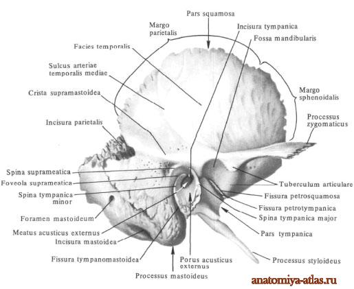
The scaly part, pars squamosa, is involved in the formation of the lateral walls of the skull. It belongs to the integumentary bones, i.e. it ossifies on the basis of connective tissue and has a relatively simple structure in the form of a vertical plate with a rounded edge overlapping the corresponding edge of the parietal bone, margo squamosa, in the form of fish scales, which is where its name comes from.
On its cerebral surface, facies cerebralis, traces of the brain, finger impressions, impressiones digitatae, and an upward groove from a. meningea media. The outer surface of the scales is smooth, participates in the formation of the temporal fossa and is therefore called facies temporalis. The zygomatic process, processus zygomaticus, departs from it, which goes forward to connect with the zygomatic bone. At its origin, the zygomatic process has two roots: anterior and posterior, between which there is a fossa for articulation with the lower jaw, fossa mandibularis. An articular tubercle, tuberculum articulare, is placed on the lower surface of the anterior root, which prevents the head of the mandible from dislocating forward when the mouth opens significantly.
The tympanic part, pars tympanica, of the temporal bone forms the anterior, lower and part of the posterior edge of the external auditory canal, ossifies endesmally and, like all integumentary bones, has the appearance of a plate, only sharply curved.
The external auditory canal, meatus acusticus extern us, is a short canal that goes inward and somewhat forward and leads into the tympanic cavity. The upper edge of its external opening, porus acusticus externus and part of the posterior edge are formed by the scales of the temporal bone, and along the remaining length by the tympanic part.
In a newborn, the external auditory canal is not yet formed, since the tympanic part is an incomplete ring (anulus tympanicus), covered by the eardrum. Due to such a close position of the eardrum outward in newborns and children early age Diseases of the tympanic cavity are more common.
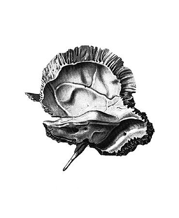
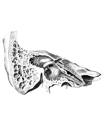
An important part of the temporal bone is the petrous part, pars petrosa, so named for the strength of its bone substance, due to the fact that this part of the bone is both involved in the base of the skull and is the bony seat of the organs of hearing and balance, which have a very delicate structure and require strong protection from damage. It develops on the basis of cartilage. The second name of this part is the pyramid, given by its shape of a trihedral pyramid, the base of which faces outward, and the apex faces forward and inward. sphenoid bone.
The pyramid has three surfaces: front, back and bottom. The anterior surface is part of the bottom of the middle cranial fossa; the posterior surface faces posteriorly and medially and forms part of the anterior wall of the posterior cranial fossa; the lower surface faces down and is visible only on the outer surface of the base of the skull. The external relief of the pyramid is complex and is determined by its structure as a container for the middle (tympanic cavity) and inner ear (bone labyrinth, consisting of the cochlea and semicircular canals), as well as the passage of nerves and blood vessels. On the anterior surface of the pyramid, near its apex, there is a noticeable small depression, impressio trigimini, from the trigeminal ganglion (n. trigeminus). Two thin grooves run outward from it, the medial one is sulcus n. petrosi majoris, and lateral-sulcus n. petrosi minoris. They lead to two foramina of the same name: the medial one, hiatus canalis n. petrosi majoris, and lateral, hiatus canalis n. re trosi minoris. Outside of these openings, an arched elevation is noticeable, etineptia arcuata, formed due to the protrusion of the rapidly developing labyrinth, in particular the superior semicircular canal. The surface of the bone between eminentia arcuata and squama temporalis forms the roof of the tympanic cavity, tegmen tympani.
Approximately in the middle of the posterior surface of the pyramid is the internal auditory opening, porus acusticus internus, which leads into the internal auditory canal, meatus acdsticus internus, where the facial and auditory nerves, as well as the internal auditory artery and veins, pass.
From the lower surface of the pyramid, facing the base of the skull, extends a thin pointed styloid process, processus styloideus, which serves as the attachment point for the muscles of the “anatomical bouquet” (mm. styloglossus, stylohyoideus, stylopharyngeus), as well as ligaments - ligg. stylohyoideum and stylomandibulare. The styloid process represents a part of the temporal bone of branchial origin. Together with lig. stylohyoideum it is a remnant of the second visceral arch, the sublingual (hyoid).
Between the styloid and mastoid processes there is a stylomastoid foramen, foramen stylomastoideum, through which n. facialis and one of the arteries enters. Medial to the styloid process there is a deep jugular fossa, fossa jugularis. Anterior to the fossa jugularis, separated from it by a sharp ridge, is the external opening of the carotid canal, foramen caroticum externum.
The pyramid has three edges: anterior, posterior and upper. The short anterior margin forms an acute angle with the scales. In this corner, the opening of the myotubal canal, canalis musculotubarius, leading into the tympanic cavity is noticeable. This channel is divided by a partition into two sections: upper and lower. The upper, smaller, semicanal, semicanalis tensoris tympani, contains this muscle, and the lower, larger, semicatialis tiibae auditvae, is the bony part of the auditory tube, which serves to conduct air from the pharynx into the tympanic cavity.
Along the upper edge of the pyramid, separating the anterior and posterior surfaces, there runs a clearly visible groove, sulcus sinus petrosi superioris, a trace of the venous sinus of the same name.
The posterior edge of the pyramid anterior to the fossa jugularis connects to the main part of the occipital bone and, together with this bone, forms the siilcus sinus petrosi inferioris - a trace of the inferior petrosal venous sinus.
The outer surface of the base of the pyramid serves as a place for muscle attachment, which determines its external relief (process, notches, roughness). Downwards it extends into the mastoid process, processus mastoideus. The sternocleidomastoid muscle is attached to it, which maintains the head in the balance necessary for an upright position of the body. Therefore, the mastoid process is absent in quadrupeds and even apes and develops only in humans in connection with their upright posture. On the medial side of the mastoid process there is a deep mastoid notch, incisura mastoidea, - the place of attachment of m. digastricus; even more inward - a small groove, sulcus a. occipitalis, - trace of the artery of the same name.
On the outer surface of the base of the mastoid process, a smooth triangle is distinguished, which is a place for rapid access to the cells of the mastoid process when they are filled with pus.
Inside the mastoid process contains these cells or cells, cellulae mastoideae, which are air cavities separated by bone bars, receiving air from the tympanic cavity, with which they communicate through the antrum mastoideum. On the cerebral surface of the base of the pyramid there is a deep groove, sulcus sinus sigmoidei, where the venous sinus of the same name lies.
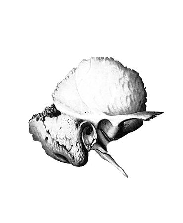
Canals of the temporal bone. The largest canal is the canalis caroticus, through which the internal carotid artery passes. Beginning with its external opening, foramen caroticum externum, on the lower surface of the pyramid, it rises upward, then bends at a right angle and opens with its internal opening, foramen caroticum internum, at the apex of the pyramid medial to the canalis musculotubarius. The canal of the facial nerve (Fig. 27), canalis facialis, begins in the depths of the porus acusticus internus, from where the canal first goes forward and laterally to the cracks (hiatus) on the anterior surface of the pyramid; at these openings the canal, remaining horizontal, turns at a right angle laterally and backwards, forming a bend - the genu, geniculum canalis facialis, and then goes down and ends through the foramen stylomastoideum, located on the lower surface of the pyramid of the temporal bone.
The organ of hearing and balance lies in the temporal bone; vessels and nerves pass through its canals. It forms the base of the skull and the lateral wall of the vault. The temporal bone articulates with the lower jaw and is the support of the masticatory apparatus.
The temporal bone develops from 6 ossification points. At the end of the second month, ossification points appear in the scaly part, in the third month - in the tympanic part, and in the fifth - in the pyramid. In the first year of life, all three parts grow together.
Structure
The temporal bone is a paired formation that has an internal and external surface. It has 3 parts located around the external auditory opening ( porus acusticus externus ): above - scaly part ( pars squamosa ), inwards and behind - the rocky part or pyramid ( pars petrosa ), in front and below - the drum part ( pars tympanica).
Scaly part
It has the shape of a plate and is located almost in the sagittal direction. Below, the scaly part is adjacent to the tympanic and petrous parts and is separated from them, respectively, by the tympanic-squamosal fissure ( fissura tympanosquamosa ) and stony-scaly(fissura petrosquamosa).
On the outer surface (in its posterior section) the groove of the middle temporal artery runs in a vertical direction ( sulcus arteriae temporalis mediae ) - trace of the junction of the artery of the same name.
Above and anteriorly from the external auditory opening, the zygomatic process extends horizontally ( processus zygomaticus ), which connects at the anterior end with the temporal process of the zygomatic bone, forming the zygomatic arch ( arcus zygomaticus ). On the anterior root of the zygomatic process there is a thickening - the articular tubercle ( tuberculum articulare ), and on the inner surface there is a postarticular tubercle ( tuberculum retroarticulare)
Between the external auditory opening and the root of the zygomatic process is the mandibular fossa ( fossa mandibularis ). It is covered with cartilage and articulates with the articular process of the lower jaw. At the bottom of this hole there is fissura petrosquamosa and fissura petrostympanica (a nerve emerges from it - the chorda tympani), which are separated by the lower process of the roof of the tympanic cavity.
The inner cerebral surface has cerebral elevations, finger-shaped depressions, and grooves for the vessels of the meninges run along it.
The scaly part has 2 edges: wedge-shaped ( margo sphenoidalis) and parietal (margo parietalis ). Accordingly, the scaly part is connected to the greater wing of the sphenoid bone and to the lower edge of the parietal bone.
Pyramid (rocky part)
It contains most of the elements of the hearing organ: the bony part of the external auditory canal, the middle and inner ear. The petrous part consists of two sections: the posterolateral - mastoid process and the anteromedial - pyramid.
Mastoid process ( Processus mastoideus) located posterior to the external auditory canal. Its outer surface is rough due to the attachment of the sternocleidomastoid muscle to it. It often has a mastoid foramen(foramen mastoideum).
The mastoid notch is visible below ( incisura mastoidea ) - the place of attachment of the posterior belly of the digastric muscle. More medially is the groove of the occipital artery ( sulcus arteria occipitalis).
On the inner surface of the brain there is a wide groove of the sigmoid sinus ( sulcus sinus sigmoidei ), at which the mastoid foramen opens ( foramen mastoideum).
Inside the mastoid process (on the cut) there are cells of different size and shape ( cellula mastoidei ). The largest of them is the mastoid cave ( antrum mastoidei ), communicating with the middle ear cavity.
The mastoid process is separated from the tympanic part by the tympanomastoid fissure ( fissura tympanomastoidea ), where the auricular branch of the vagus nerve passes ( ramus auricularis n. vagus).
Pyramidhas 3 surfaces: front, back and bottom; and, accordingly, 3 edges: top, back and front.
Front surface ( facies anterior) facing the cranial cavity. In its middle there is an arched elevation ( eminentia arcuata ), formed by the underlying semicircular canal of the labyrinth of the inner ear. Between fissura petrosquamosa and eminentia arcuata The roof of the tympanic cavity is located ( tegmentum tympani ), under which is the tympanic cavity. Near the apex of the pyramid there is a trigeminal impression ( impressio trigemeni ) - the site of contact of the trigeminal nerve ganglion. (this is a collection of nerve cells; 3 branches extend from it - n. ophthalmicus, n. maxillarisAndn. mandibularis)
The grooves of the greater and lesser petrosal sinuses extend from the apex of the pyramid ( sulcus nervi petrosi majoris et minoris ), which end in clefts of the same name ( hiatus canalis nervi petrosi majoris et minoris ). The corresponding nerves exit through these clefts. Along the upper edge of the pyramid there is a groove of the superior petrosal sinus ( sulcus sinus petrosi superior)
The back surface of the pyramid ( facies posterior) faces the cranial cavity and passes into the mastoid process. Almost in the middle of this surface is the internal auditory opening ( porus acusticus internus ), passing into the internal auditory canal ( meatus acusticus internus ). It contains the front ( VII), intermediate(XIII ), vestibulocochlear nerves ( VIII ); artery and vein of the labyrinth.
Lateral porus acusticus internus there is a subarc fossa ( fossa subarcuata ) - it includes a process of the dura mater. Even more lateral is the external aperture of the vestibule aqueduct ( apertura externa aquaeductus vestibuli ) - through it the endolymphotic duct leaves the ear cavity.
Bottom surface ( facies inferior) lies on the lower surface of the base of the skull. It contains a round or oval-shaped jugular fossa ( fossa jugularis ) - the place of contact of the superior bulb of the internal jugular vein. At the bottom of this hole there is a small groove (in it lies ramus auricularis n. vagus ), which leads into the opening of the mastoid tubule ( canaliculus mastoideus).
Anterior to the jugular fossa lies o round shape opening leading to the carotid canal ( canalis caroticus).
Between the jugular fossa and the external opening of the carotid canal there is a stony dimple ( fossula petrosa ) - place of contact of the glossopharyngeal nerve ( IX ). And in the depths of this dimple there is a hole leading into the tympanic canaliculus, where the inferior tympanic artery and tympanic nerve pass (or Jacobson's nerve - mixed (sensitive and autonomic), preganglionic, parasympathetic, coming from nucl. salivatoriusinferiorand central processes of the lower node, i.e. under for. jugulare).
Lateral from fossa jugularis there is a styloid process ( processus styloideus ), from which muscles and ligaments begin. Behind it lies the stylomastoid foramen ( foramen stylomastoideus ), which is the outlet of the facial canal.
Upper edge pyramids- sulcus sinus petrosi superior.
Rear edge pyramids- sulcus sinus petrosi inferior, near which or on which the external aperture of the cochlear tubule lies (apertura externa canaliculi cochleae)
Anterior edge of pyramid - fissura petrosquamosa , on which the opening of the myotubal canal is located lateral to the internal opening of the carotid canal.
Drum part
This is the smallest section of the temporal bone, which forms the anterior, inferior and part of the posterior wall meatus acusticus externus and limits porus acusticus externus . The tympanic cavity is covered with a mucous membrane and contains 3 auditory ossicles: the malleus, the incus and the stapes.
Temporal bone apertures:
1) Apertura externa aquaeductus vestibuli
2) Apertura externa canaliculi cochlea
3) Apertura superior canaliculi tympani - corresponds hiatus canalis nervi petrosi minoris.
4) Apertura inferior canaliculi tympani - lies at the bottom fossula petrosa.
Temporal bone canals
1) Carotid canal (Canalis caroticus ) - begins on the lower surface of the pyramid with an external hole, then, bending almost at a right angle, opens at the top of the pyramid with an internal hole. The internal carotid artery passes through this canal.
2) Carotid tympanic tubules ( Canaliculi caroticotympanici ) - pass through the wall of the carotid canal near its external opening and open into the tympanic cavity. The carotid-tympanic nerves and arteries pass through here.
3) Facial canal (Canalis (nervi) facialis ) - begins at the bottom of the internal auditory canal, goes forward and laterally to the level of the cleft of the canal of the greater petrosal nerve. Here the channel forms the first bend - the elbow ( geniculum canalis facialis ). Next, the canal goes laterally and backward, follows the axis of the stony part and at the level eminentia pyramidalis forms the second limb of the facial canal. And opens in foramen stylomastoideum . The facial nerve passes through this canal ( VII pair of cranial nerves)
4) Drum string channel ( Canaliculus chorda tympani ) - is a branch from the facial canal and begins slightly higher foramen stylomastoideum . It enters the tympanic cavity and ends in fissura petrotympanica . A branch of the facial nerve, the chorda tympani, passes through this canal.
5) Tympanic canaliculus ( Canaliculus tympanicus ) - starts in a pertura inferior canaliculi tympani , enters the tympanic cavity, passes along its medial wall in the groove of the promontory ( sulcus promontorii ), follows to the top wall and opens into a pertura superior canaliculi tympani . This canal contains the lesser petrosal nerve and a branch of the glossopharyngeal nerve - Jacobson's nerve.
6) Musculo-tubal canal (with analis muculotubarius ) - the external opening of the canal is located in the notch between the scaly and petrous parts and runs almost along the axis of the pyramid. A horizontally located septum divides the canal into two semi-canaliculi: semicanalis musculi tensoris tympani (upper and smaller, it contains the muscle that strains the tympanic membrane) and semicanalis tubae auditivae (lower and larger, the Eustachian tube passes through it, which connects the tympanic cavity with the pharyngeal cavity)
7) Mastoid tubule ( Canaliculus mastoideus ) - starts at the bottom fossa jugularis , crosses the lower part of the facial canal and opens into fissura tympanomastoideus . The canal contains the auricular branch of the vagus nerve ( X pair of cranial nerves).
Project Correspondent EstheticLife"
Serova Ksenia
Temporal bone
Encyclopedia "Anatomy watch online. Human anatomy in pictures"
Temporal bone
os temporale , steam room, participates in the formation of the base of the skull and the side wall of its vault. It contains the organ of hearing and balance. It articulates with the lower jaw and is the support of the masticatory apparatus.On the outer surface of the bone there is an external auditory opening, porus acusticus externus, around which three parts of the temporal bone are located: above - the scaly part, pars squamosa, inwardly and behind - the stony part (pyramid), pars petrosa, in front and below - the tympanic part, pars tympanica.
The scaly part, pars squamosa, has the shape of a plate and is located almost in the sagittal direction. The outer temporal surface, facies temporalis, of the scales is slightly rough and slightly convex. In the posterior section, the groove of the middle temporal artery, sulcus arteriae temporalis mediae (trace of the junction of the artery of the same name), runs in a vertical direction.
In the posteroinferior part of the squamosal part there is an arcuate line that continues into the lower temporal line of the parietal bone.
From the scaly part, above and somewhat anterior to the external auditory opening, the zygomatic process, processus zygomaticus, extends horizontally. Beginning with a wide root, the zygomatic process then narrows. It has an inner and outer surface and two edges - an upper, longer one, and a lower, shorter one. The anterior end of the zygomatic process is jagged and connects with the temporal process, processus temporalis, of the zygomatic bone, forming with it the zygomatic arch, arcus zygomaticus.
On the lower surface of the root there is a transversely oval-shaped articular mandibular fossa, fossa mandibularis, into which the head of the mandible articulates with it enters. Anteriorly, the articular fossa is limited by the articular tubercle, tuberculum articulare.
The outer surface of the scaly part is involved in the formation of the temporal fossa, fossa temporalis (bundles of the temporal muscle, m. temporalis, begin here). The inner, cerebral, surface of the scales, facies cerebralis, is slightly concave. It has digital impressions, impressiones digitatae, and cerebral elevations, as well as an arterial groove, sulcus arteriosus (it contains the middle meningeal artery, a. meningea media).
The squamous part of the temporal bone has two free edges - the sphenoid and the parietal.
The anteroinferior, wedge-shaped edge, margo sphenoidalis, is wide, serrated, connects with the scaly edge of the large wing of the sphenoid bone and forms a wedge-squamosal suture, sutura sphenosquamosa. The upper posterior, parietal edge, margo parietalis, is pointed, longer than the previous one, connected to the scaly edge of the parietal bone. The petrous part, pars petrosa (pyramid), of the temporal bone consists of posterolateral and anteromedial sections.
The posterolateral part of the petrous part of the temporal bone is the mastoid process, processus masloideus, which is located posterior to the external auditory opening; it distinguishes between outer and inner surfaces. The outer surface is convex, rough and is the site of muscle attachment. Inferiorly, the mastoid process turns into a cone-shaped protrusion, which can be easily felt through the skin.
On the inside, the process is limited by the deep mastoid notch, incisura mastoidea (the posterior belly of the digastric muscle, venter posterior m. digastrieus, originates from it). Parallel to the notch and somewhat posteriorly there is a groove of the occipital artery, sulcus arteriae occipitalis (trace of the junction of the artery of the same name). At the base of the mastoid process, on its outer surface, there is often a mastoid opening, foramen mastoideum; sometimes it is in the seam. connecting the mastoid process with the occipital bone, and belongs to the group
venous graduates.
On the inner, cerebral, surface of the mastoid process there is a wide S-shaped groove of the sigmoid-Impresslo trigemiiil prominent sinus, sulcus sinus sigmoidei, passing at the top into the groove of the same name of the parietal bone and further into the groove of the transverse sinus of the occipital bone (it contains the venous sinus of the dura mater). shells). Downwards, the groove of the sigmoid sinus continues into the groove of the same name in the occipital bone.
At the back, the mastoid process is bounded by the jagged occipital edge, margo occipitalis, which, connecting with the mastoid edge of the occipital bone, forms the occipital-mastoid suture. sutura occipitomastoidea. In this suture, in the middle of its length or in the occipital edge, there is a mastoid opening, foramen mastoideum (sometimes there are several of them), which, as already mentioned, is the location of the mastoid emissary veins, vv. emissariae mastoideae, connecting the saphenous veins of the head with the sigmoid venous sinus, and the mastoid branch of the occipital artery, ramu smastoi-deus a. occipitalis. From above, the mastoid process is limited by the parietal edge, which, at the border with the same edge of the scales of the temporal bone, forms the parietal notch, incisura parietalis; the mastoid angle of the parietal bone enters here, forming the parietomastoid suture, sutura parietomastoidea.
At the place of transition of the outer surface of the mastoid process into the outer surface of the scales, one can notice the remains of the squamous-mastoid suture, sutura squamosomasoidea, which is clearly visible on the skull of children.
A cut of the mastoid process reveals the bony air cavities inside it - mastoid cells, cellulae mastoideae, separated from one another by bone walls. The permanent cavity is the mastoid cave, antrum mastoideum, located in the central part of the process; The mastoid cells open into it, and it connects with the tympanic cavity. The mastoid cells and mastoid cave are lined with mucous membrane.
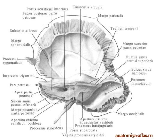
The anteromedial part of the petrous part lies medial to the squamosal part and the mastoid process. It has the shape of a triangular pyramid, the long axis of which is directed outward and behind and forward. The base of the stony part is directed outward and posteriorly; apex of the pyramid, apex partis petrosae. directed inward and anteriorly.
In the stony part there are three surfaces: anterior, posterior and lower, and three edges: upper, posterior and anterior.
The anterior surface of the pyramid, facies anterior partis pertosae, faces the cranial cavity, is smooth and wide, is directed obliquely from top to bottom and forward and passes into the cerebral surface of the scaly part, from which it is sometimes separated by a stony-squamous fissure, fissura petrosquamosa. Almost in the middle of the anterior surface there is an arcuate elevation, eminentia arcuata, which is formed by the anterior semicircular canal of the labyrinth underlying it. Between the elevation and the stony-scaly fissure there is a small platform - the roof of the tympanic cavity, legmen tympani. under which is the tympanic cavity, cavum tym-pani. On the anterior surface near the apex of the petrous part there is a small trigeminal depression, impressio tri-gemini (place of contact of the trigeminal nerve ganglion).
Lateral to the depression there is a cleft of the canal of the greater petrosal nerve, hiatus canalis nervi pelrosi majoris. from which a narrow groove of the greater petrosal nerve, sulcus nervi petrosimajoris, extends medially. Anterior and somewhat lateral to this opening there is a small cleft of the canal of the lesser petrosi nerve, hiatus canalis nervi petrosi minoris, from which the groove of the lesser petrosi nerve, sulcus nervi petrosi minoris, is directed.
Posterior surface of the pyramid, facies posterior partis petrosa e , like the anterior surface, faces the cranial cavity, but is directed upward and posteriorly, where it passes into the mastoid process. Almost in the middle of it there is a rounded internal auditory opening, porus acusticus internus, which leads to the internal auditory canal, meatus acusticus internus (it contains the facial, intermediate, pre-door cochlear nerves, as well as the artery and vein of the labyrinth, a. et v. labirinthi). A little higher and lateral from the internal auditory opening there is a subarcuate fossa, well-defined in newborns of small depth, fossa subarcuata (it includes a process of the dura mater). Also lateral from the internal auditory opening lies the slit-shaped external aperture of the vestibule aqueduct, apertwa externa aquaeductus vestibuli (through The endolymphatic duct emerges from the cavity of the inner ear). The lower surface of the pyramid, facies inferior partis petrosae, is rough and uneven, lying on the lower surface of the base of the skull. On it there is a round or oval-shaped jugular fossa, fossa jugularis (the location of the superior bulb of the internal jugular vein). At the bottom of this fossa there is a small groove (the auricular branch of the vagus nerve passes through it). The groove leads into the opening of the mastoid canaliculus. canaliculus mastoideus, which opens in the tympanomasoid fissure, fissura tym-panomastoidea. The posterior edge of the jugular fossa is limited by the jugular notch, incisura jugul a ris, which is a small intrajugular process, processus intrajugularis. divides into two parts: anteromedial and posterolateral. Anterior to the jugular fossa there is a rounded hole; it leads to the sleepy canal. canalis caroticus, opening at the top of the stony part with a hole.
Between the anterior circumference of the jugular fossa and the external opening of the carotid canal there is a small stony dimple, fossula petrosa (place of contact of the lower ganglion of the glossopharyngeal nerve). In the depths of the dimple there is a hole that opens the passage into the tympanic canaliculus, canaliculus tympanicus (the tympanic nerve and the inferior tympanic artery pass through it). The tympanum leads into the middle ear, auris media, or the tympanic cavity, cavum tympani.
Laterally from the jugular fossa, a styloid process, processus styloideus, directed downwards and slightly anteriorly of varying length, protrudes. from which muscles and ligaments begin.
In front and outside the root of the process, the bony protrusion of the tympanic part descends - the vagina of the styloid process, vagina processus slyloidei.
Behind the root of the process there is an awl-mastoid opening, foramen stylomastoideum, which is the exit opening of the facial canal, canalis facialis.
The upper edge of the pyramid, margo superior partis petrosae, separates its front surface from the back. Along the edge there is a groove of the superior petrosal sinus, sulcus sinus petrosi superioris - an imprint of the superior petrosal venous sinus lying here and the attachment of the tentorium cerebellum - part of the dura mater. This groove passes posteriorly into the groove of the sigmoid sinus of the mastoid process of the temporal bone.
The posterior edge of the pyramid, margo posterior partis petrosae, separates its posterior surface from the lower one. Along it, on the brain surface, runs the groove of the inferior stony sinus, sulcus sinus petrosi inferioris (trace of the contact of the inferior stony venous sinus). Almost in the middle of the posterior edge, near the jugular notch, there is a triangular funnel-shaped depression in which lies the external aperture of the cochlear canaliculus, apertura externa canaliculi cochleae.
The anterior edge of the petrous part, located on the lateral side of its anterior surface, is shorter than the upper and posterior ones; it is separated from the squamous part of the temporal bone by a petrosquamous fissure, fisswa petrosquamatosa. On it, lateral to the internal opening of the carotid canal, there is the opening of the muscular-tubal canal, canalis
musculolubarius, leading into the tympanic cavity (see Musculo-tubal canal).


899 rub

Sports anatomy
It explains in an intelligible form how the human body works and how its individual parts interact with each other, how bones, muscles, tendons and ligaments behave under different loads. Drawings and photographs clearly illustrate the topics discussed and allow readers to look inside their own bodies. For wide range readers.
479 rub
Human Anatomy 360?. Illustrated atlas
COMPLETE 3D GUIDE This book provides detailed 3D models of every structure in the human body. An innovative method of body modeling provides a unique opportunity to see with your own eyes previously inaccessible areas of the body. Modern technologies make it possible to demonstrate the thinnest sections of tissues, organs and cells and show the three-dimensional structure of different parts of the body from all sides. Carefully checked texts supplemented the latest facts about the achievements of medicine. Composite spine and stereo vario cover.
1739 rub
Muscles. Anatomy. Movements. Testing
This book, prepared by a group of German specialists working in the field of rehabilitation, became a bestseller in the West, going through 5 editions. It is a reference book on the muscles of the human body, aimed at diagnosing disorders of their function.
Each muscle is considered depending on its specific joint and axes of movement. In each case, synergists and antagonists of all possible muscle movements are given. This structure of the book allows you to immediately obtain the entire amount of information on the muscle.
The book is well illustrated. Particular emphasis was placed on the superficial anatomy of the musculoskeletal system to facilitate orientation in muscle topography, identification and palpation.
For orthopedists, neurologists, rheumatologists, physical therapy doctors, physiotherapists, manual therapists, occupational therapists, massage therapists and other specialists involved in musculoskeletal system, his diseases, treatment and rehabilitation.
1779 rub

Guide to the body. A practical guide to body palpation
The book "Guide to the Body" is a unique guide to palpation of the musculoskeletal system. In the structure of this book, areas of the body and movements in the body are considered according to the logic of their interaction: shoulder girdle and shoulder, forearm and hand, spine and torso, head, neck and face, pelvis and thigh, lower leg and foot. Each area is described in detail, from superficial anatomy, skin and fascia, to muscles and bones.
In total, the book describes 162 muscles, 206 bones, 33 ligaments, and 110 bony landmarks. The total number of illustrations is about 1400. The Appendix provides valuable information on synergistic muscles, facial muscles and a guide to trigger points.
For chiropractors, osteopaths, massage specialists, physiotherapists, physical therapy.
The book is intended for specialists in the field of obstetrics and gynecology.
539 rub

Lower limb. Functional anatomy. Volume 2
There is no longer any need to introduce Dr. Adalbert I. Kapandji, world famous in the circles of orthopedic surgeons and kinesitherapists. After a long career as an orthopedic surgeon, later engaged in hand surgery, he devoted his time entirely to the re-edition and updating of the three volumes of his work "Functional Anatomy" (previously the work was called "Physiology of the Joints." The second volume of this work is devoted to lower limb and sheds light on such complex concepts as the heterokinetic cardan of the ankles and bones of the hindfoot, the concept of the lower leg. The book also includes a summary chart of knee stability factors, a table of lower limb nerves, and a new chapter on the physiology of gait. As in previous editions, Dr. Adalbert I. Kapanji independently prepared and colored all the diagrams and drawings to help better understand human biomechanics. To date, the book has been translated into 11 languages.... ... In the Atlas, along with drawings traditionally showing appearance various organs, there are pictures of the macro-micro- and microscopic structure of organs and tissues, as well as text, diagrams and tables accompanying the pictures, which help to better understand and evaluate the structure and design of the organs of the human body. A special feature of the atlas is its numerous, in comparison with other publications of this kind, complex topographic images blood vessels and nerves. Plastic three-dimensional diagrams of the structure of organs, for example, the liver, spleen, lung, kidneys, organ of Corti, etc., are very useful.
The first volume is dedicated skeletal system, joint system, muscular system....
3742 rub
Os temporale - steam room, complex in shape and structure. It participates in the formation of the base of the skull and complements the lateral walls of the roof of the skull. The organs of hearing and balance are located in the temporal bone; nerves and blood vessels pass through its canals. On its outer surface there is the external auditory opening, porus acusticus extemus, around which there are three parts of the temporal bone: the squamous bone, pars squamosa; stony, pars petrosa and drum, pars tympanica.
The scales and tympanic part develop on the basis of connective tissue, and the stony part develops on the basis of cartilage.
Scaly part, pars squamosa, has the shape of a thin plate located in the sagittal plane. A squamoid suture connects its free edge with the lower edge of the parietal bone and the greater wing. The lower part of the scales is adjacent to the stony and tympanic part and is delimited from it by a stony-scaly fissure, fissura petrosquamosa, and from the tympanic part by a tympanic-squamous fissure, fissura thympanosquamosa. In the posterior part of the scales there is a groove of the middle temporal artery, sul. A. temporalis mediae. In the posterior-inferior section, the temporal line is distinguished. Above the scales and slightly forward, the zygomatic process, processus zygomaticus, extends, which originates with a wide root and then tapers. It fuses with the process of the zygomatic bone and forms the zygomatic arch, arcus zygomaticus. On the lower surface of the root there is an articular mandibular fossa, fossa mandibularis. In front, the articular fossa is limited by the articular tubercle, tuberculum articulare, and behind by the extraglobal tubercle, tuberculum retroarticulare. The inner medullary surface of the squama contains finger-like indentations, cerebral protrusions and grooves from the middle meningeal artery, a. meningea media, in case of damage to the scales, aneurysms of this artery may occur; this should be taken into account in clinical (neurosurgical) practice.
Rocky part, pars petrosa, has the shape of a triangular pyramid, the top of which is directed inward and forward, and the base is directed back and sideways. In the stony part, the following surfaces are distinguished: anterior, fades anterior partis petrosae, posterior, fades posterior partis petrosae, and lower, fades inferior partis petrosae, as well as the upper, posterior and anterior corner.
The front surface of the pyramid faces the cranial cavity. Almost in the middle of the anterior surface there is an arcuate elevation, eminentia arcuata, which coincides with the anterior semicircular canal of the labyrinth.
The roof of the tympanic cavity is adjacent to the elevation. On the anterior surface of the pyramid, at its apex, there is a trigeminal squeeze, impressio trigeminalis, for the trigeminal ganglion. To the side of it is the formation of the canal of the greater petrosal nerve, hiatus canalis n. petrosi majoris, from which the medial groove of the greater petrosal nerve, sul. n. petrosi majoris. Slightly in front and to the side is the upper course of the lesser petrosal nerve, hiatus canalis n. petrosi minoris, from which the groove of the lesser petrosal nerve, sul. n. petrosi minoris. The nerves of the same name exit through these holes.
On the posterior surface of the stony part, almost in the middle, there is an internal auditory opening, porus acusticus internus, leading to the internal auditory canal, meatus acusticus internus. At the upper edge of the stony part, in the area between the internal auditory opening and the external opening of the aqueduct of the vestibule, there is a subarc fossa, fossa subarcuata. And at the lower edge there is the opening of the cochlea's aqueduct, apertura externa agueductus cochleae. Above and to the side of this opening is the external opening of the vestibular aqueduct, apertura externa agueductus vestibuli, through which the endolymphatic duct, ductus endolymphaticus, passes.
On the lower surface of the stony part there is an oval-shaped jugular fossa, fossa jugularis, at the bottom of which there is a groove leading to the opening of the mastoid canaliculus. The posterior edge of the jugular fossa is limited by the jugular notch, incisura jugularis. Anterior to the jugular fossa is the external opening of the carotid canal, apertura externa canalis carotid, which leads into the carotid canal, canalis caroticus, which opens at the apex of the petrous part with an internal opening, apertura interna canalis carotid. At the external opening on the posterior wall of the carotid artery canal there are openings of the carotid-tympanic tubules, canaliculi caroticotympanici, opening into the tympanic cavity through which vessels and nerves pass. Between the jugular fossa and the external opening of the carotid canal there is a stony dimple, fossula petrosa, in the depth of which is the lower opening of the tympanic tubule, apertura inferior canaliculi tympanici (BNA). On the side of the jugular fossa there is a downward styloid process, processus styloideus, which is the site of attachment of the “anatomical bouquet” (mm. styloglossus, stylohyoideus, stylophmyngeus) and the ligament ligg. stylohyoideum et stylomandibular. Behind the root of the process there is a stylomastoid opening, foramen stylomastoideus. In front and outside the styloid process there is a bony protrusion of the tympanic part - the vagina of the styloid process, vagina processus styloidei.
The upper edge of the stony part separates its anterior surface from the posterior one. Along this edge runs the upper stony groove, sul. sinus petrosi superioris. The posterior edge of the petrous portion separates the posterior surface from the inferior surface. Along this edge runs the lower stony groove, sul. sinus petrosi inferioris. The anterior edge of the stony part separates its anterior surface from the lower one. On it, on the side of the internal opening of the carotid canal, there is an opening of the muscular-tubal canal, canalis musculotubarius, which connects the tympanic cavity with the nasal part of the pharynx.
Below, the base of the stony part is elongated into the mastoid process, processus mastoideus, the outer surface on it is rough from the muscle attached to it. At the opening of the mastoid process, cells, cellulae mastoidei, are visible, lined with mucous membrane. The largest cell, called the mastoid cave, antrum mastoideum, communicates with the cavity of the middle ear. In the case of inflammation of the middle ear (otitis), the infection can penetrate into the cells and lead to purulent inflammation (mastoiditis), the treatment of which requires surgery.
Externally, two grooves run from the mastoid process: the medial one - for the occipital artery, sul. a. occipitalis, and slightly to the side - the mastoid notch, incisura mastoidea. The mastoid process is separated from the tympanic part by the tympanomastoid fissure, fissura tympanomastoidea, which is the site of passage of the auricular branch of the vagus nerve.
In the area between the occipital bone and the mastoid process there is a mastoid foramen, the foramen mastoideum is the widest. Basically, the mastoid foramen is located in the occipital-mastoid suture (C. Libersa, 1934). On the outer surface of the mastoid process, the mastoid triangle (Shipo) is conventionally distinguished, which is the site of trephination (anthrotomy) for inflammatory conditions of the middle ear (cells and cave). On the inner surface of the mastoid process there is a groove of the sigmoid sinus, sul. sinus sigmoidei. Along with the S-shaped form, there are hook-shaped, sickle-shaped, straight and arched forms (G.D. Burdei, 1951, 1955). In the middle section of the groove of the sigmoid sinus, the mastoid opening, foramen mastoideum, opens, through which the mastoid emissary vein passes, connecting the sigmoid sinus with the suboccipital venous plexus.
Drum part, pars tympanica, located around the external auditory canal, meatus acusticus extemus. It limits below and behind the external auditory opening, porus acusticus extemus, and the tympanic cavity, cavitas tympanica, and with its free edge it fuses with the scales and the mastoid process.
It is separated from the scales by a tympanic-squamous fissure, fissura tympanosquamosa, into which a process of the roof of the tympanic cavity is inserted. It divides it into two fissures: the stony-squamous, fissura petrosquamosa, and the stony-tympanic, fissura petrotympanica (Glaseri), through which a branch of the intermediate nerve emerges from the tympanic cavity - the chorda tympani. Above the external auditory opening is the suprachnoid spine, spina suprameatica. The cartilaginous part of the auditory canal is attached to the free rough edge of the tympanic part, which limits the external auditory opening.
Ossification. A baby's temporal bone is made up of three parts. The first ossification points appear in the scales at 8 weeks intrauterine development, and for 3 months - in the drum part. At the 5th month, five points of ossification appear in the cartilaginous base of the petrous part. In a newborn, parts of the temporal bone are separated by gaps filled with connective tissue, which grow together during the first year of life.
Canals and cavities of the temporal bone
There are seven canals of the temporal bone: 1. Facial nerve canal, canalis n. facialis;2. Musculo-tubal canal, canalis musculotubarius;
3. Carotid canal, canalis caroticus
4. Carotid-tympanic tubules, canaliculi caroticotympanic;
5. Canaliculus chorde tympanv,
6. Tympanic tubule, canaliculus tympanicus;
7. Mastoid tubule, canaliculus mastoideus.
Facial nerve canal, canalis n. facialis - originates at the bottom of the auditory canal, goes at a right angle to the axis of the petrous part and goes to the formation of the large petrosal nerve, hiatus canalis n. petrosi majoris, where it turns and forms the elbow of the facial canal, geniculum canalis facialis. The ganglion of the geniculate facial nerve is located here, from which the greater petrosal nerve arises. After which it passes along the posterior wall of the tympanic cavity, forming protrusions of the facial canal, prominentia canalis facialis, then the canal goes vertically downwards, where it opens with a stylomastoid opening, foramen stylomastoideus. The canal contains the facial and intermediate nerves (VII pair), the superficial petrosal branch from the middle meningeal artery and the stylomastoid artery and vein.
Musculo-tubal canal, canalis musculotubarius - originates in the notch between the petrous and squamosal parts of the temporal bone and runs along the axis of the petrous part. The bony septum divides it into two semi-canals: the upper - the semi-canal of the muscle that stretches the tympanic membrane, semicanalis musculi tensoris tympani, and the lower - the semi-canal of the auditory tube, semicanalis tube auditoriae (LNA). The upper one contains a muscle that stretches the eardrum, the lower one connects the tympanic cavity with the pharyngeal cavity.
Sleepy channel, canalis caroticus - originates on the lower surface of the stony part with the external carotid foramen, apertura externa canalis carotici. The canal goes upward, passes in front of the tympanic cavity, forming a bend, and then goes forward and medially, opening with the internal carotid foramen, apertura interna canalis carotid, at the apex of the petrous part. The canal contains the internal carotid artery, the veins that accompany it, and a nice nerve plexus.
Carotid tympanic tubules, canaliculi caroticotympanici - small tubules that branch from the carotid canal and lead into the tympanic cavity. The carotid-tympanic nerves pass through here.
Drum String Canadian, canaliculus chorde tympani - originates on the wall of the facial canal above the stylomastoid foramen, goes forward and upward, enters the tympanic cavity and opens on its posterior wall. A branch of the intermediate nerve passes through the canal - the chorda tympani, which exits the tympanic cavity through the petrotympanic fissure.
Tympanic canaliculus, canaliculus tympanicus - originates in the petrosal fossa, fossula petrosa, then through the lower wall enters the tympanic cavity, passes along its medial wall, and rises upward, where it opens with the gap of the lesser petrosal nerve, hiatus canalis n. petrosi minoris. The tympanic nerve passes through the canal, which, at the exit from the tympanic cavity, is called the lesser petrosal nerve (branch of the IX pair).
Mastoid tubule, canaliculus mastoideus - originates in the depths of the jugular fossa, crosses the facial canal in its lower part and opens in the tympanomastoid fissure. The auricular branch of the vagus nerve passes through the canal.
Tympanic cavity, cavitas tympania, discussed in the “Sense Organs” section.
Os temporale, steam room, is involved in the formation of the base of the skull and the lateral wall of its vault. It contains the organ of hearing and balance. It articulates with and is the support of the masticatory apparatus.
On the outer surface of the bone there is an external auditory opening, porus acusticus externus, around which three parts of the temporal bone are located; on top is the scaly part, inwardly and behind is the stony part, or pyramid, in front and below is the tympanic part.
The scaly part, pars squamosa, has the shape of a plate and is located almost in the sagittal direction. The outer temporal surface, facies temporalis, of the scaly part is slightly rough and slightly convex. In the posterior section, the groove of the middle temporal artery, sulcus arteriae temporalis mediae (trace of the junction of the artery of the same name), runs in a vertical direction.
In the posterior lower part of the scaly part there is an arcuate line, which continues into the lower temporal line, linea temporalis inferior,.
From the scaly part, above and somewhat anterior to the external auditory opening, the zygomatic process, processus zygomaticus, extends horizontally. It is, as it were, a continuation of the supramastoid crest, crista supramastoidea, located horizontally along the lower edge of the outer surface of the scaly part. Beginning with a wide root, the zygomatic process then narrows. It has an inner and outer surface and two edges - a longer upper one and a shorter lower one. The anterior end of the zygomatic process is serrated. Zygomatic process of the temporal bone and temporal process, processus temporalis, the zygomatic bones are connected using the temporomygomatic suture, sutura temporozygomatica forming the zygomatic arch, arcus zygomaticus.
On the lower surface of the root of the zygomatic process there is a transverse oval-shaped mandibular fossa, fossa mandibularis. The anterior half of the fossa, up to the petrosquamosal fissure, is the articular surface, fades articularis, of the temporomandibular joint. Anteriorly, the mandibular fossa is limited by the articular tubercle, tuberculum articulare.
The outer surface of the scaly part is involved in the formation of the temporal fossa,
fossa temporalis (beams begin here, m. temporalis).
The inner cerebral surface, facies cerebralis, is slightly concave. It has finger-like impressions, impressiones digitatae, as well as an arterial groove, sulcus arteriosus (it contains the middle meningeal artery, a. meningea media).
The squamous part of the temporal bone has two free edges - the sphenoid and the parietal.
The anteroinferior sphenoid edge, margo sphenoidalis, is wide, serrated, connects with the scaly edge of the large wing of the sphenoid bone and forms a sphenoid-squamosal suture, sutura sphenosquamosa.
The superior posterior parietal edge, margo parietalis, is pointed, longer than the previous one, connected to the scaly edge of the parietal bone.
The pyramid (stony part), pars petrosa, of the temporal bone consists of posterolateral and anteromedial sections.

The posterolateral part of the petrous part of the temporal bone is the mastoid process, processus mastoideus, which is located posterior to the external auditory opening. It distinguishes between outer and inner surfaces. The outer surface is convex, rough and is the site of muscle attachment. Inferiorly, the mastoid process passes into a cone-shaped protrusion, which can be easily felt through the skin,
On the inside, the process is limited by the deep mastoid notch, incisura mastoidea (the posterior belly of the digastric muscle, venter posterior m. digastrici, originates from it). Parallel to the notch and somewhat posteriorly there is a groove of the occipital artery, sulcus arteriae occipitalis (trace of the junction of the artery of the same name).
On the inner, medullary, surface of the mastoid process there is a wide S-shaped groove of the sigmoid sinus, sulcus sinus sigmoidei, which passes at the top into the groove of the same name of the parietal bone and further into the groove of the transverse sinus of the occipital bone (it contains the venous sinus, sinus transversa). Downwards, the groove of the sigmoid sinus continues as the groove of the same name of the occipital bone.
The posterior border of the mastoid process is the jagged occipital edge, margo occipitalis, which, connecting with the mastoid edge of the occipital bone, forms the occipital-mastoid suture, sutura occipitomastoidea. In the middle of the length of the suture or in the occipital edge there is a mastoid foramen, foramen mastoideum (sometimes there are several of them), which is the location of the mastoid veins, vv. emissariae mastoidea, connecting the saphenous veins of the head with the sigmoid venous sinus, as well as the mastoid branch of the occipital artery, ramus mastoideus a. occipitalis.
From above, the mastoid process is limited by the parietal edge, which, at the border with the same edge of the squamous part of the temporal bone, forms the parietal notch, incisura parietalis; the mastoid angle of the parietal bone enters it, forming the parietomastoid suture, sutura parietomastoidea.
At the place of transition of the outer surface of the mastoid process into the outer surface of the scaly part, one can notice the remains of the squamous-mastoid suture, sutura squamosomastoidea, which is clearly visible on the skull of children.
On the cut of the mastoid process, the bony air cavities located inside it are visible - mastoid cells, cellulae mastoideae. These cells are separated from one another by the bony mastoid walls, paries mastoideus. The permanent cavity is the mastoid cave, antrum mastoideum, in the central part of the process; The mastoid cells open into it, it connects with the tympanic cavity, cavitas tympanica. The mastoid cells and mastoid cave are lined with mucous membrane.
The anteromedial part of the petrous part lies medial to the squamosal part and the mastoid process. It has the shape of a triangular pyramid, the long axis of which is directed from the outside and from behind to the front and medially. The base of the stony part is directed outward and posteriorly; the apex of the pyramid, apex partis petrosae, is directed inward and anteriorly.
In the stony part there are three surfaces: anterior, posterior and lower, and three edges: upper, posterior and anterior.
The anterior surface of the pyramid, facies anterior partis petrosae, is smooth and wide, facing the cranial cavity, directed obliquely from top to bottom and forward and passes into the brain surface of the scaly part. It is sometimes separated from the latter by a stony-scaly fissure, fissura petrosquamosa. Almost in the middle of the anterior surface there is an arcuate elevation, eminentia arcuata, which is formed by the anterior semicircular canal of the labyrinth underlying it. Between the elevation and the stony-scaly fissure there is a small platform - the roof of the tympanic cavity, tegmen tympani, under which is the tympanic cavity, cavum tympani. On the anterior surface, near the apex of the petrous part, there is a small trigeminal depression, impressio trigemini (attachment site of the trigeminal ganglion, ganglion trigeminale).
Lateral to the depression is the cleft of the canal of the greater petrosal nerve, hiatus canalis n. petrosi majoris, from which a narrow groove of the greater petrosal nerve, sulcus n., extends medially. petrosi majoris. Anterior and somewhat lateral to this opening there is a small cleft of the canal of the lesser petrosal nerve, hiatus canalis n. petrosi minoris, from which the groove of the lesser petrosal nerve, sulcus n., is directed. petrosi minoris.
The posterior surface of the pyramid, facies posterior partis petrosae, like the anterior one, faces the cranial cavity, but is directed upward and posteriorly, where it passes into the mastoid process. Almost in the middle of it there is a round-shaped internal auditory opening, porus acusticus internus, which leads into the internal auditory canal, meatus acusticus internus (facial, intermediate, vestibular-cochlear nerves, nn. facialis, intermedius, vestibulocochlearis, as well as an artery pass through it and vein of the labyrinth, a. et v. labirinthi). A little higher and lateral from the internal auditory opening there is a well-defined subarcuate fossa (fossa subarcuata) in newborns, of small depth (it includes a process of the dura mater of the brain). Even more lateral lies the slit-like external aperture of the vestibule aqueduct, apertura externa aqueductus vestibuli, which opens into the vestibule aqueduct, aqueductus vestibuli. The endolymphatic duct emerges from the cavity of the inner ear through the aperture.
The lower surface of the pyramid, facies inferior partis petrosae, rough and uneven, forms part of the lower surface of the base of the skull. On it there is a round or oval jugular fossa, fossa jugularis (the location of the superior bulb of the internal jugular vein).
You might be interested in this read: