Sphenoid bone, os sphenoidale , unpaired, forms the central section of the base of the skull.
middle part sphenoid bone- body, corpus, cubic shape, has six surfaces. On the upper surface, facing the cranial cavity, there is a recess - the Turkish saddle, sella turcica, in the center of which is the pituitary fossa, fossa hypophysialis. It contains the pituitary gland, hypophysis. The size of the fossa depends on the size of the pituitary gland. The border of the Turkish saddle in front is the tubercle of the saddle, tuberculum sellae. Behind it, on the lateral surface of the saddle, there is an unstable middle inclined process, processus clinoideus medius.
Body of the sphenoid bone– corpus sphenoidalis
pituitary fossa-fossahypophysialis
saddle tubercle–tuberculumsellae
Posterior inclined processes processus clinoidei posterioris
Anterior inclined process–processus clinoideusanterior
Carotid furrow-sulcuscaroticum
wedge-shaped uvula– lingual sphenoidalis
wedge-shaped beak-rostrum sphenoidale
wedge-shaped shell-conchae sphenoidalis
Sphenoid sinus aperture– aperture sinus sphenoidalis
Sphenoid sinus– sinus sphenoidalis
small wing– ala minor
big wing–ala major
visual channel-canalis opticus
Superior orbital fissure– fissura orbitalis superior
round hole-foramen rotundum
oval hole–foramenovale
spinous foramen–foramenspinosus
cerebral surface-facies cerebralis
finger-like impressions–impressionsdigitales
arterial sulcus-sulcusarteriosi
Orbital surface–faciesorbitales
Maxillary surface–faciesmaxillaries
Temporal surface–faciestemporalis
Infratemporal ridge-cristainfratemporalis
pterygoid process–processuspterygoideus
pterygoid canal-canalispterygoideus
Sphenoid spine– spinaossis sphenoidalis
Medial plate–laminamedialis
Lateral plate–laminalateralis
pterygoid fossa–fossapterygoidea
Pterygoid notch–incisurapterygoidea
Pterygoid hook-hamulus pterygoideas
Body of the sphenoid bone
On the upper surface of the body there is a depression - the Turkish saddle, containing the pituitary gland. The anterior border of the saddle is the tubercle of the saddle, the posterior border is the back of the saddle. On the sides of the Turkish saddle are carotid grooves with cavernous sinuses, in which the internal carotid arteries and associated nerve plexuses pass. Anterior to the tubercle of the saddle is the chiasm furrow, on which the optic chiasm is located. The back of the saddle protrudes forward in the lateral sections, forming the posterior inclined processes. The back surface of the back of the Turkish saddle smoothly continues with the upper surface of the basilar part of the occipital bone, forming a slope.
In front, the body of the sphenoid bone is connected to the perpendicular plate of the ethmoid bone and the vomer through a vertically located wedge-shaped ridge. Posteriorly, the body of the sphenoid bone fuses with the basilar part of the occipital bone.
Most of the body of the sphenoid bone is made by the airy sphenoid sinus, divided by a septum into two halves. In front, the sinus is limited by wedge-shaped shells located on the sides of the wedge-shaped crest. The shells form holes - apertures through which the wedge-shaped cavity communicates with the nasal cavity. The walls of the sphenoid sinus are lined with a mucous membrane.
small wings
The lesser wings are directed away from the anteroposterior corners of the body in the form of two horizontal plates. At their base there are rounded holes, which are the beginning of the visual canals containing the optic nerves and ophthalmic arteries. The upper surfaces of the lesser wings face the cranial cavity, the lower surfaces face the cavity of the orbits, forming the upper walls of the upper orbital fissures. The front edges of the wings articulate with the orbital parts of the frontal bone. The posterior margins lie freely in the cranial cavity, being the border of the anterior and middle cranial fossae.
Small wings are connected to each other by a wedge-shaped elevation located in front of the decussation furrow.
Big wings
Large wings extend outward from the lateral surfaces of the body of the bone. The large wing has four surfaces and three edges. At the base of the large wing there are three openings: a round opening through which the maxillary nerve passes; oval, through which the mandibular nerve passes; spinous (it passes the middle meningeal artery, vein and nerve).
Large wing surfaces
The medulla, superior, faces the cranial cavity.
The orbital surface, anteroposterior, has a rhomboid shape. It is turned into the cavity of the orbit, forming part of its lateral wall. The lower edge of the orbital surface of the wing, along with the posterior edge of the orbital surface of the upper jaw, forms the inferior orbital fissure.
The maxillary surface, anterior, has a triangular shape, small size. From above it is limited by the orbital surface, from the side from below - by the root of the pterygoid process. The maxillary surface is involved in the formation of the posterior wall of the pterygopalatine fossa. It has a round hole.
The temporal surface, superior lateral, is divided by the infratemporal crest into the directly temporal and pterygoid surfaces. The temporal surface is involved in the formation of the temporal fossa. Oval and spinous openings open on the pterygoid surface. The pterygoid surface forms the anterior wall of the infratemporal fossa.
The edges of the big wing
The frontal edge, upper, is connected to the orbital part of the frontal bone by means of a wedge-frontal suture. The outer sections of the frontal edge end with a sharp parietal edge, forming a wedge-parietal suture with the parietal bone. The internal sections of the frontal margin pass into a thin free margin, which limits the superior orbital fissure from below.
The zygomatic edge, anterior, connects to the frontal process of the zygomatic bone, forming the sphenoid-zygomatic suture.
Scaly edge, posterior, connects with wedge-shaped edge temporal bone and forms a wedge-scaly suture. Behind and outside, the scaly edge ends with the spine of the sphenoid bone. Inward from the spine, the scaly edge is located in front of the stony part of the temporal bone, forming with it a wedge-stony gap, passing medially into a torn hole.
pterygoid processes
Pterygoid processes (lat. processus pterygoidei) begin at the junction of the large wings with the body of the sphenoid bone and are located vertically downwards. At the base of the processes are pterygoid canals, in which the nerves and vessels of the same name pass. Anteriorly, each canal opens into the pterygopalatine fossa.
Each process consists of medial and lateral plates, which are fused in the anterior-upper sections, limiting the pterygoid fossa in front. The free, unfused ends of the plates limit the pterygoid notch filled with the pyramidal process of the palatine bone. The lower end of the medial plate ends with a pterygoid hook directed downward and outward.
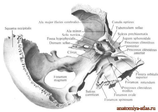
Occipital bone, os occipitale, forms the posterior and lower walls of the cranium, participating simultaneously in the vault of the skull and at its base. Accordingly, it (being a mixed bone) ossifies as an integumentary bone on the soil of the connective tissue (scales of the occipital bone), as well as on the soil of cartilage (the rest of the bone). In humans, it is the result of the fusion of several bones that exist independently in some animals. Therefore, it consists of 4 parts that are laid down separately, fusing into a single bone only at the age of 3-6 years. These parts that close big hole occipital bone, foramen magnum(the place where the spinal cord passes into the oblong from the spinal canal into the cranial cavity), the following: in front - the main part, pars basilaris(in animals - os basilare), on the sides - side parts, partes latrales(in animals - ossa lateralia), and behind - the occipital scales, squama occipitalis(in animals - os superius). Top part the scales, wedged between the parietal bones, ossify separately and often remain separated by a transverse suture for life, which is also a reflection of the existence in some animals of an independent interparietal bone, os interparietal, as it is called in humans.
Scales of the occipital bone, squama occipitalis, as the integumentary bone has the form of a plate, convex on the outside and concave on the inside. Its external relief is due to the attachment of muscles and ligaments. So, in the center of the outer surface is external occipital protuberance, protuberdntia occipitalis externa(place of appearance of the ossification nucleus). From the hillock laterally goes on each side along a curved line - upper line, tinea nuchae superior. A little higher there is a less noticeable one - linea nuchae suprema (the highest). From the occiput down to the posterior edge of the foramen magnum runs along the midline external occipital crest, crista occipitalis externa. From the middle of the ridge to the sides go lower you are other lines, lineae nuchae inferiores. The relief of the inner surface is due to the shape of the brain and the attachment of its membranes, as a result of which this surface is divided by means of two ridges crossing at right angles into four pits; both of these ridges together form cruciform eminence, emineniia cruciformis, and at the place of their intersection - internal occipital protuberance, protuberdntia occipitalis interna. The lower half of the longitudinal ridge is sharper and is called crista occipitalis interna, while the upper and both halves (often right) of the transverse ridge are provided with well-defined grooves: sagittal, sulcus sinus sagittdlis superioris, and transverse, sulcus sinus transversi (traces of adjoining venous sinuses of the same name).
Each of side parts, partes laterales, participates in the connection of the skull with the spine, therefore, on its lower surface it carries occipital condyle, condylus occipitalis- place of articulation with the atlas. Approximately about the middle of the condylus occipitalis passes through the bone canal of the hypoglossal nerve, candlis (nervi) hypoglossi.
On the upper surface of the pars lateralis is sulcus sinus sigmoidei (a trace of the venous sinus of the same name).
Main part, pars basilaris, by the age of 18 fuses with the sphenoid bone, forming a single bone in the center of the base of the skull os basilare. On the upper surface of this bone there is a slope, merged from two parts, clivus, on which lies the medulla oblongata. On the lower surface, which is part of the upper wall of the pharynx, protrudes pharyngeal tubercle, tuberculum pharyngeum to which the fibrous membrane of the pharynx is attached.
Sphenoid bone
Sphenoid bone, os sphenoidale, unpaired, resembles a flying insect, which is the reason for the name of its parts (wings, pterygoid processes). Therefore, the term "wedge-shaped" is unfortunate and appeared, apparently, by mistake *. It is also called the main one, since it is located in the center of the base of the skull.
* (In Galen's manuscript, this bone was called sphecoidal (similar to a wasp), but its scribe is believed to have made a mistake and wrote sphenoid (sphenoid).)
The sphenoid bone is the product of the fusion of several bones that exist independently in animals, therefore it develops as a mixed bone from several paired and unpaired ossification foci, which merge into 3 parts by the time of birth, which in turn fuse into a single bone by the end of the first year of life. It distinguishes the following parts: 1) the body, corpus(in animals - unpaired basisphenoid and presphenoid); 2) big wings, alae majores(in animals - paired alisphenoid); 3) small wings, alae minores(in animals - paired orbitosphenoid), and 4) pterygoid processes, processus pterygofdei(its medial plate, a former paired pterygoid, develops on the basis of connective tissue, while all other parts of the bone arise on the basis of cartilage).
Body, corpus, on its upper surface has a recess along the midline - Turkish saddle, sella turcica, at the bottom of which lies a fossa for an appendage of the brain, fossa hypophysidlis. In front of it is an elevation, tuberculum sellae, along which the sulcus chiasmatis passes transversely for the decussation (chiasma *) of the optic nerves; at the ends of the sulcus chiasmatis are visible visual channels, candles optici through which the optic nerves pass from the cavity of the orbit to the cavity of the skull. Behind the Turkish saddle is limited to the bone plate, back Turkish saddle, dorsum sellae. On the lateral surface of the body runs a curved carotid groove, sulcus caroticus, a trace of the internal carotid artery.
* (The correct pronunciation is chiasma, chiasmatis.)
On the anterior surface of the body, which is part of the posterior wall of the nasal cavity, a crest, crista sphenoidlis, is visible below, entering between the wings of the vomer. Crista sphenoidalis articulates anteriorly with the perpendicular plate of the ethmoid bone. On the sides of the scallop, irregularly shaped holes are visible, aperturae sinus sphenoiddlis, leading to the airway sinus, sinus sphenoiddlis, which is located in the body of the sphenoid bone and is divided by a septum, septum sinuum sphenoiddlium, into two halves. Through these openings, the sinus communicates with the nasal cavity.
In a newborn, the sinus is very small and only around the 7th year of life begins to grow rapidly.
small wings, alae minores, are two flat triangular-shaped plates, which extend forward and laterally from the anterior superior edge of the body of the sphenoid bone with two roots; between the roots of the small wings are the mentioned visual canals, canales optici. Between the small and large wings is superior orbital fissure, fissura orbitalis superior leading from the cavity of the skull to the cavity of the orbit.
Big wings, alae majores, depart from the lateral surfaces of the body laterally and upward. Near the body, posterior to the fissura orbitalis superior, there is round hole, foramen rotundum, leading anteriorly to the pterygopalatine fossa, due to the passage of the second branch of the trigeminal nerve, n. trigemini. Posteriorly, a large wing in the form of an acute angle protrudes between the scales and the pyramid of the temporal bone. Near it there is spinous foramen, foramen spinosum, through which passes a. meningea media. Much more is visible in front of him. foramen ovale, through which the third branch n passes. trigemini.
Large wings have four surfaces: brain, fades cerebrdlis, orbital, fdcies orbitdlis, temporal, fades temporalis, And maxillary, fdcies maxilldris. The names of the surfaces indicate the areas of the skull where they face. The last two surfaces are separated by a ridge, crista infratempordlis.
pterygoid processes, processus pterygoidei, depart from the junction of the large wings with the body of the sphenoid bone vertically down. Their base is pierced by a sagittally running canal, candlis pterygoideus, - the place where the named nerve and vessels pass. The anterior opening of the canal opens into the pterygopalatine fossa.
Each process consists of two plates - lamina medidlis and lamina lateralis, between which a pit is formed behind, fossa pterygoidea.
The medial plate below is folded with a hook, hamulus pterygoideus, through which the tendon of m. tensor veli palatini (one of the muscles of the soft palate).
Temporal bone
Temporal bone, os tempordle, a paired bone, has a complex structure, since it performs all 3 functions of the skeleton and not only forms part of the side wall and base of the skull, but also contains the organs of hearing and balance. It is the product of the fusion of several bones (mixed bone) that exist independently in some animals, and therefore consists of three parts: 1) the scaly part, pars squamosa(in animals - os squamosum); 2) drum part, pars tympanica(in animals - tympanicum), and 3) the stony part, pars petrosa(in animals - petrosum).
During the 1st year of life, they merge into a single bone, closing the external auditory canal, meatus acusticus externus, in such a way that the scaly part lies above it, the stony part is medially from it, and the drum part is behind, below and in front. Traces of the fusion of individual parts of the temporal bone are preserved for life in the form of intermediate sutures and fissures, namely: on the border of pars squamosa and pars petrosa, on the anterior upper surface of the latter - fissura petrosquamosa; in the depths of the jaw fossa - fissura tympanosquamosa, which is divided by the process of the stony part into fissura petrosquamosa and fissura petrotympdnica (the chorda tympani nerve exits through it).
scaly part, pars squamosa, participates in the formation of the lateral walls of the skull. It belongs to the integumentary bones, that is, it ossifies on the soil of the connective tissue and has a relatively simple structure in the form of a vertically standing plate with a rounded edge superimposed on the corresponding edge of the parietal bone, margo squamosa, in the form of fish scales, hence its name.
On cerebral surface her, fdcies cerebrdlis, traces of the brain are visible, finger impressions, impressiones digitdtae, and an ascending groove from a. meningea media. The outer surface of the scales is smooth, participates in the formation of the temporal fossa and is therefore called fdcies temporalis. Departs from her zygomatic process, processus zygomdticus, which goes forward at the connection with the zygomatic bone. At its beginning, the zygomatic process has two roots: anterior and posterior, between which there is a fossa for articulation with the lower jaw, fossa, mandibularis. On the lower surface of the anterior root is placed articular tubercle, tuberculum articuldre, preventing dislocation of the head of the lower jaw forward with a significant opening of the mouth.
drum part, pars tympanica, the temporal bone forms the anterior, lower and part of the posterior edge of the external auditory canal, ossifies endesmally and, like all integumentary bones, has the form of a plate, only sharply curved.
External auditory canal, meatus acusticus externus, is a short channel heading inward and somewhat forward and leading into the tympanic cavity. The upper edge of its outer opening, porus acusticus externus, and part of the posterior edge are formed by the scales of the temporal bone, and the rest of the length by the tympanic part.
Newborn the external auditory canal has not yet been formed, since the tympanic part is an incomplete ring (anulus tympanicus), tightened by the tympanic membrane. Due to such a close location of the tympanic membrane outward in newborns and children early age diseases of the tympanic cavity are more often observed.
An important part of the temporal bone is rocky part, pars petrosa, so named for the strength of its bone substance, due to the fact that this part of the bone simultaneously participates in the base of the skull, and is the bone receptacle of the hearing and balance organs, which have a very thin structure and need strong protection from damage. It develops on the basis of cartilage. The second name of this part is the pyramid, given by its shape of a trihedral pyramid, the base of which is turned outward, and the top is forward and inward to the sphenoid bone.
The pyramid has three surfaces; front, back and bottom. The anterior surface is part of the bottom of the middle cranial fossa; the back surface is turned back and medially and forms part of the anterior wall of the posterior cranial fossa; the lower surface is turned downwards and is visible only on the outer surface of the base of the skull. The external relief of the pyramid is complex and is due to its structure as a receptacle for the middle (tympanic cavity) and inner ear (a bony labyrinth consisting of the cochlea and semicircular canals), as well as the passage of nerves and blood vessels. On front surface pyramids, near its apex, there is a noticeable slight depression, impressio trigemini, from the trigeminal ganglion (n. trigeminus). Two thin grooves pass outward from it, the medial one is sulcus n. petrosi majoris, and lateral - sulcus n. petrosi minoris. They lead to two openings of the same name: medial, hiatus candlis n. petrosi majoris, and lateral, hiatus candlis n. petrosi minoris. Outside of these holes is noticeable arcuate eminence, eminentia arcudta, formed due to the protrusion of a rapidly developing labyrinth, in particular the upper semicircular canal. The surface of the bone between the eminentia arcuata and the squama temporalis forms the roof of the tympanic cavity, tegmen tympani.
Approximately in the middle rear surface the pyramid is internal auditory opening, porus acusticus internus, which leads to internal auditory canal, meatus acusticus internus where the facial and auditory nerves pass, as well as the internal auditory artery and veins.
From bottom surface pyramid facing the base of the skull, a thin pointed styloid process, processus styloideus, serving as a place of attachment of the muscles of the "anatomical bouquet" (mm. styloglossus, stylohyoideus, stylopharyngeus), as well as ligaments - ligg. stylohyoideum and stylomandibular. The styloid process is part of the temporal bone of branchial origin. Together with lig. stylohyoideum, it is a remnant of the second visceral arch, the hyoid (hyoid).
Between the styloid and mastoid processes is awl mastoid foramen, foramen stylomastoideum, through which n exits. facialis and one of the arteries enters. Medially from the styloid process is a deep jugular fossa, fossa juguldris. Anterior to the fossa jugularis, separated from it by a sharp ridge, is the external opening of the carotid canal, foramen caroticum externum.
The pyramid has three edges: front, back and top. The short anterior margin forms an acute angle with the scales. There is a hole in this corner. musculotubular canal, canalis musculotubdrius leading to the tympanic cavity. This channel is divided by a partition into two sections: upper and lower. Upper, smaller, semi-canal, semicanalis m. tensoris tympani, contains this muscle, and the lower, larger, semicanalis tubae auditivae, is the bone part of the auditory tube, which serves to conduct air from the pharynx into the tympanic cavity.
On the upper face of the pyramid, which separates the anterior and posterior surfaces, there is a clearly visible groove, sulcus sinus petrosi superidris, a trace of the venous sinus of the same name.
The rear edge of the pyramid anterior to the fossa jugularis connects to the main part of the occipital bone and forms, together with this bone, sulcus sinus petrosi inferioris - a trace of the lower petrosal venous sinus.
The outer surface of the base of the pyramid serves as a place of muscle attachment, which is the reason for its outer relief (process, notches, roughness). From top to bottom, it stretches into mastoid process, processus mastoideus. The sternocleidomastoid muscle is attached to it, which maintains the head in balance, necessary for the vertical position of the body. Therefore, the mastoid process is absent in tetrapods and even anthropoid apes and develops only in humans in connection with his upright posture. On the medial side of the mastoid process there is a deep mastoid notch, incisdra mastoidea, - place of attachment m. digastricus; even more inwards - a small furrow, sulcus a. occipitalis, - a trace of the artery of the same name.
On the outer surface of the base of the mastoid process, a smooth triangle is isolated, which is a place for quick access to the cells of the mastoid process when they are filled with pus.
Inside the mastoid process and contains these cells or cells, cellulae mastoideae, which are air cavities separated by bone bars, receiving air from the tympanic cavity, with which they communicate through the antrum mastoideum. On the cerebral surface of the base of the pyramid there is a deep groove, sulcus sinus sigmoidei, where the venous sinus of the same name lies.
Canals of the temporal bone. The largest channel is canalis caroticus through which the internal carotid artery passes. Starting with its outer opening, foramen caroticum externum, on the lower surface of the pyramid, it rises upward, then bends at a right angle and opens with its internal opening, foramen caroticum internum, at the top of the pyramid medially from canalis musculotubarius. facial nerve canal(fig. 27), canalis facialis, begins in the depths of the porus acusticus internus, from where the canal first goes forward and laterally to the cracks (hiatus) on the front surface of the pyramid; at these openings, the canal, remaining horizontal, turns at a right angle laterally and backward, forming a bend - the knee, geniculum canalis facialis, and then goes down and ends through the foramen stylomastoideum, located on the lower surface of the temporal bone pyramid.
Parietal bone
Parietal bone, os parietale, steam room, forms the middle part of the cranial vault. In man, it reaches the greatest development in comparison with all animals in connection with highest development he has a brain. It represents a typical integumentary bone, which performs mainly the function of protection. Therefore, it has a relatively simple structure in the form of a quadrangular plate, convex on the outside and concave on the inside. Its four edges serve to connect with neighboring bones, namely: the front - with the frontal, margo frontalis, back - from the occipital, margo occipitalis, upper - with the same name bone of the other side, margo sagittalis, and lower - with scales of the temporal bone, margo squamosus. The first three edges are serrated, and the last is adapted to form a scaly suture. Of the four corners, the anterior superior one connects to the frontal bone, angulus frontalis, anteroinferior with sphenoid bone, angulus sphenoidalis, posterior superior with occipital bone, angulus occipitalis, and posterior with the base of the mastoid process of the temporal bone, angulus mastofdeus. The relief of the outer convex surface is due to the attachment of muscles and fascia. At its center stands parietal tubercle, tuber parietale(place of ossification). Below it are curved temporal lines - lineae tempordles (superior et inferior) - for the temporal fascia and muscle. Near the upper edge there is a hole, foramen parietale (for the artery and venous outlet). The relief of the inner concave surface, fdcies interna, is due to the fit of the brain and especially the dura mater; the places of attachment of the latter to the bone have the form of a sagittal sulcus, sulcus sinus sagittalis superioris (trace of the venous sinus, sinus sagittalis superior), and also in the region of angulus mastoideus of the transverse sulcus, sulcus sinus sigmoidei (trace of the venous sinus of the same name). The vessels of this membrane seemed to be imprinted in the form of grooves branching almost on the entire inner surface. On the sides of the sulcus sinus sagittalis superioris, traces of the so-called pachyonic granulations, foveolae granuldres, are visible (see the meninges).
frontal bone
Frontal bone, os frontale, unpaired, participates in the formation of the cranial vault and refers to its integumentary bones, developing on the basis of connective tissue. In addition, it is associated with the senses (smell and vision). According to this dual function, it consists of two sections: vertical - scales, squama frontalis, and horizontal. The latter, according to the relation to the organs of vision and smell, is divided into a paired orbital part, pars orbitalis, and unpaired nasal, pars nasalis. As a result, 4 parts are distinguished in the frontal bone:
1. Scales, squama frontalis, like any integumentary bone, has the form of a plate, convex on the outside and concave on the inside. It ossifies from two points of ossification, noticeable even in an adult on outer surface, fades externa, in the form of two frontal tubercles, tubera frontdlia. These bumps are expressed only in humans in connection with the development of the brain. They are absent not only in great apes, but even in extinct forms of man. The lower edge of the scales is called the supraorbital, mdrgo supraorbitalis. Approximately on the border between the inner and middle thirds of this region, there is supraorbital notch, incisura supraorbitalis(sometimes turns into foramen supraorbital), the place of passage of the arteries and nerve of the same name. Immediately above the supraorbital margin, prominently varying in size and extent of the eminence is noticeable - brow ridges, arcus superciliares, which medially in the middle line pass into a more or less prominent platform, glabella (glabella). It is a reference point when comparing the skulls of modern man with a fossil. The outer end of the supraorbital margin extends into zygomatic process, processus zygomatics connecting with the zygomatic bone. From this process goes up a clearly visible temporal line, llnea temporalis, which limits temporal surface scales, facies temporalis. On inner surface, facies interna, along the midline there is a groove from the posterior edge, sulcus sinus sagittdlis superioris, which passes into frontal scallop, crista frontalis. These formations are the attachment of the dura mater. Near the midline, pits of pachyon granulations (outgrowths of the arachnoid membrane of the brain) are noticeable.
2 and 3. Orbital parts, partes orbitae, represent two horizontally located plates, which, with their lower concave surface, face the orbit, the upper one - into the cranial cavity, and are connected to the sphenoid bone with their posterior edge. On the upper cerebral surface there are traces of the brain - impressiones digitatae. Inferior surface, facies orbitlis, forms the upper wall of the orbit and bears traces of adhering eye accessories; at the zygomatic process - pit of the lacrimal gland, fossa gldndulae lacrimdlis, near the incisura supraorbitalis - fovea trochledris and a small spike, spina trochledris, where a cartilaginous block (trochlea) is attached to the tendon of one of the muscles of the eye. Both orbital parts are separated from each other by a notch, incisura ethmoidlis, which is filled on the whole skull with an ethmoid bone.
4. bow, pars nasalis, occupies the anterior part of the lattice notch along the midline; here a scallop is noticeable, which ends in a sharp process - spina nasalis, which takes part in the formation of the nasal septum. On the sides of the scallop are pits that serve as the upper wall for the cells of the ethmoid bone; anterior to them there is a hole leading to frontal sinus, sinus frontalis, - a cavity that is located in the thickness of the bone behind the superciliary arches and the size of which varies greatly. The frontal sinus, which contains air, is usually divided by a septum, septum sinuum frontdlium. In some cases, there are additional frontal sinuses behind or between the main ones (Jovanovic, 1961). The frontal bone is the most characteristic of all the bones of the skull for humans. In the most ancient hominids (as well as great apes), it was sharply tilted back, forming a sloping, "running back" forehead. Behind the orbital constriction, it sharply divided into scales and orbital parts. Along the edge of the eye sockets, from one zygomatic process to the other, a continuous thick ridge ran. In modern man, the roller has sharply decreased, so that only the superciliary arches remain of it. According to the development of the brain, the scales straightened and took a vertical position, at the same time the frontal tubercles developed, as a result of which the forehead from sloping became convex, giving the skull a characteristic appearance.
Ethmoid bone
Ethmoid bone, os ethmoidale, unpaired, is usually described among the bones of the brain skull, although for the most part it participates in the formation of the facial skull. Located centrally between the bones of the face, it comes into contact with most of them, participating in the formation of the nasal cavity and eye sockets, and is closed by them on the whole skull. It develops in connection with the nasal capsule, on the basis of cartilage, built from thin bone plates surrounding the air cavities (Fig. 28). The bone plates of the ethmoid bone are located in the form of the letter "T", in which the vertical line is perpendicular plate, lamina perpendicularis, and the horizontal lattice plate, lamina cribrosa. Hanging from the latter on the sides of the lamina perpendicularis lattice labyrinths, labyrfnthi ethmoidales. As a result, 4 parts can be distinguished in the ethmoid bone:
1. Lamina cribrosa- a rectangular plate that performs incisura ethmoidalis of the frontal bone. It is pierced like a sieve with small holes (hence its name), through which branches of the olfactory nerve (about 30) pass. On its midline rises cockscomb, crista gdlli(place of attachment of the dura mater).
2. Lamina perpendicularis is part of the nasal septum.
3 and 4. Labyrfnthi ethmoidales represent a paired complex of bone air cells, cellulae ethmoidales, covered from the outside by a thin orbital plate, lamina orbitdlis, which forms the medial wall of the orbit (Fig. 29). The upper edge of the orbital plate is connected to the orbital part of the frontal bone, anteriorly - with the lacrimal bone, behind - with the sphenoid and orbital process of the palatine, from below - with the upper jaw; all these bones cover the marginal cellulae ethmoidales. On the medial side of the labyrinths are two turbinates - conchae nasdles superior et media, although there is sometimes a third - concha nasalis suprema.
sinks represent curved bone plates, due to which the surface of the nasal mucosa covering them increases.
The sphenoid bone is a large bone element of the skull, formed by the fusion of several bones. The articulations form the central section of the base of the skull: the side walls, part of the brain and facial sections.
In the structure of the skeleton, there are several more bones with the same name - the triclinoid bones of the foot. Included in the bone structure of the midfoot.
cranial bone
The anatomy here is complex, including the body and three paired elements: the greater wing, the lesser wing, and the pterygoid process.
The body of the sphenoid bone is cubic, with a sinus inside. The structure is determined by six functional surfaces: top, back, front, bottom and two side.
The body connects with the occipital, ethmoid bone of the skull, the orbital processes of the palatine bone, the wings of the vomer, and the orbital plates. The sides pass into small and large wings. At the top there is a recess for the location of the pituitary gland. Pass through the body:
- optic nerve;
- carotid and basilar arteries;
- medulla;
- bridge.
Anatomy of small wings. Plates with roots, between which there is a canal with the optic nerve. Anteriorly, the wings form a serrated junction with the frontal and ethmoid bones of the skull. The back smooth edge does not connect to anything. The dura mater is attached to the inclined processes.
The upper surface of the small wing faces the cranial cavity, and the lower surface is involved in the formation of the walls of the orbit. The cavity between the small and large wing is called the superior orbital fissure, several nerves pass there.
Anatomy of the great wing. Wide base with three holes. The 2nd and 3rd branches of the trigeminal nerve pass through the round and oval. The spinous foramen is small, through it runs the middle meningeal artery. The large wing has four surfaces: cerebral, maxillary, temporal, orbital.
The pterygoid process extends vertically down from the base of the greater wing. Vessels and nerves lie in the narrow pterygoid canal. The anterior edge of the process extends to the pterygopalatine fossa, the posterior edge to the outer base of the skull in the region of the sphenoid spine.
It has medial and lateral plates fused in front. The second is wider and shorter. The posterior edge of the plates diverges into the pterygoid fossa, the lower edge is notched. The medial downward passes into the pterygoid hook.
Bone damage
The sphenoid bone of the skull structure has a complex structure. She is involved in the formation of many departments of the cranium. Nerves run through it blood vessels. All this plus the proximity of the brain make her fracture very dangerous for the life of the victim.
Any head injury is considered quite a serious cause for concern for the health and life of the patient. Even if there is no fracture, the brain, blood vessels, nerves or internal organs can be damaged.
Violation of the integrity of the bone tissue is classified as a fracture of the base of the skull. It can be an independent injury or be accompanied by a fracture of the arch.
The severity of the injury is determined by the number of damaged elements. A fracture with a displacement is more dangerous; nearby tissues and organs can be injured.
The complex of treatment is selected based on the nature of the injury and the existing complications. Antibacterial prophylaxis is required, as well as sanitation of the nasal and ear cavities, ophthalmological, neurological, surgical and ENT diagnostics.
Conservative treatment is provided in case of non-severe injuries, surgical if present:
- comminuted fracture;
- compression or injury to the brain;
- liquorrhea or purulent infections.
The anatomy of the foot is not as complex as in the skull. The sphenoid bone is not one here, there are three of them. They are located in front of the scaphoid. They are part of the midfoot.
Articularly connected to each other and the metatarsal bones of the foot. The intermediate cuneiform bone is somewhat shorter than the other two.
The wide side of the intermediate and lateral faces upwards, while the third bone faces downwards. The posterior articular areas form the navicular articulation. There are also articular platforms on the sides of contact with each other and at the junctions with other elements of the foot.
Bone damage
A midfoot fracture is rare. You can only get such an injury as a result of a direct blow or a fall of a heavy object.

By nature, it is more often a fracture without displacement or comminuted. Complicated by torn ligaments. Located at the inner edge of the foot, the medial bone is more susceptible to external impact, but this does not exclude the fracture of all three bones.
During treatment, local anesthesia, arch modeling, immobilization for 1.5 months are performed. After that, a complex of rehabilitation and preventive measures is required.
As you can see, the described elements of the skeleton, apart from the name, have little in common. But knowing the location of the bone, as well as taking into account the peculiarities of its structure, it is possible to foresee the consequences of the injuries received.
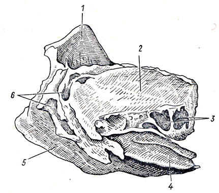
- Pterygoid process, processus pterygoideus. Rice. A, B.
- Lateral plate [pterygoid process] lamina lateralis. Rice. A, B.
- Medial plate [pterygoid process], lamina medialis. Rice. A, B.
- Pterygoid notch, incisura pterygoidea. It is located between the two plates of the pterygoid process and is directed downward. It is filled with the pyramidal process of os palatinum. Rice. A.
- Pterygoid fossa, fossa pterygoidea. It is located between the lateral and medial plastics. Place of attachment m.pterygoideus medialis. Rice. A, B.
- Navicular fossa, fossa scaphoidea. An indentation at the base of the medial plate of the pterygoid process. Starting place mjensor veli palatini. Rice. A.
- Vaginal process, processus vaginalis. It is located on the inner side of the base of the medial plate of the pterygoid process. Rice. A, B.
- Palato-vaginal sulcus, sulcus palatovaginal. Together with the palatine bone, it forms the canal of the same name. Rice. B.
- Vaginal sulcus, sulcus vomerovaginal. It is located at the base of the pterygoid process and, together with the vomer, forms the canal of the same name. Rice. B.
- Pterygoid hook, hamulus pterygoideus. It is located at the end of the medial plate of the pterygoid process and is directed downward. Rice. A, B.
- Furrow pterygoid hook, sulcus hamuli pterygoidei. Formed by a sharp bend of the pterygoid hook. Rice. B.
- Pterygoid [[vidian]] canal, canalis pterygoideus []. Passes at the base of the pterygoid process towards the pterygopalatine fossa. Contains large and deep stony nerves Fig. A. See fig. IN.
- Pterygoid-spinous process, processus pterygospinosus. Sharp projection on the posterior margin of the lateral plate of the pterygoid process. Rice. A.
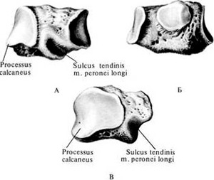
- Temporal bone, os temporale. It is located between the occipital, sphenoid and parietal bones. Consists of stony, tympanic and scaly parts. Rice. B, G, D.
- Pyramid (rocky part), pars petrosa. Contains the organ of hearing and balance. Rice. G.
- Occipital margin, margo occipitalis. Connects to the occipital bone. Rice. V, G.
- Mastoid process, processus mastoideus. Located behind the external auditory canal. Rice. V, D.
- Mastoid notch, incisura mastoidea. It is located on the lower surface of the pyramid, medially from the mastoid process. Place of the beginning of the posterior abdomen m.digastricus. Rice. IN.
- Groove of the sigmoid sinus, sulcus sinus sigmoidei. Rice. G.
- Furrow of the occipital artery, sulcus a.occipitalis. It is located at the occipital edge of the pyramid, the medial mastoid notch. Rice. IN.
- mastoid opening, foramen mastoideum. Located behind the mastoid process. Contains the emissary vein. Rice. V, G.
- Facial canal, canalis facialis. It begins in the internal auditory canal and ends with the stylomastoid foramen. Contains the nerve of the same name. Rice. B, G, D.
- Ring of the facial canal, geniculum canalis facialis. The bend of the facial canal at the anterior wall of the pyramid, near the cleft of the large stony nerve. Rice. G.
- Drum string tubule, canaliculus chordae tympani. A narrow passage connecting the facial canal and the tympanic cavity. Contains drum string. Rice. G, D.
- Top of the pyramid, apex partis petrosae. Directed forward and medially. Rice. V, G.
- Sleepy canal, canalis caroticus. It begins on the outer base of the skull between the jugular foramen and the musculo-tubal canal. Contains the internal carotid artery. Rice. IN.
- Carotid tympanic tubules, canaliculi caroticotympanic. Pass in the wall of the carotid canal. They contain vessels and nerves that lead to the tympanic cavity. Rice. IN.
- Musculo-tubal canal, canalis musculotubarius. It is located in front of the carotid canal and leads to the tympanic cavity. Contains the auditory tube and the muscle that strains the eardrum. Rice. V, D.
- The semi-canal of the muscle that strains the eardrum, semicanalis m.tensoris tympani. Rice. D.
- Semicanal of the auditory tube, semicanalis tubae auditoriae (auditivae). Rice. D.
- Septum of the muscular-tubal canal, septum canalis musculotubarii. Bone wall, between the semi-canals mentioned above. Rice. D.
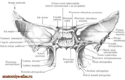
Body of the sphenoid bone corpus ossis sphenoidalis, the middle part of the bone, cubic in shape, has six surfaces. The upper surface of the body, facing the cranial cavity, has a recess in its middle sections - the Turkish saddle, sella turcica. in the center of which is the pituitary fossa. It contains the pituitary gland. The size of the fossa is determined by the size of the pituitary gland. The pituitary fossa is especially vulnerable in case of premature birth. The fusion of the two nuclei of ossification of the fossa occurs on the 8th month of intrauterine life. This raises the possibility of damage to the structure of the pituitary fossa with subsequent dysfunction of the pituitary gland. The Turkish saddle is limited in front by the tubercle of the saddle, tuberculum sellae. Behind it, on the lateral surface of the saddle, there is a non-permanent middle inclined process, processus clinoideus medius. Anterior to the tubercle of the saddle there is a shallow transverse furrow of the decussation, sulcus chiasmatis. On it lies the optic chiasm, chiasma opticum. On the sides, the furrow passes into the optic canal, canalis opticus. Ahead of the furrow is a smooth surface - a wedge-shaped elevation, jugum sphenoidale connecting the small wings of the sphenoid bone. The anterior edge of the upper surface of the body is serrated, protrudes slightly forward and connects with the posterior edge of the perforated plate, lamina cribrosa, ethmoid bone, forming a wedge-ethmoid suture, sutura sphenoethmoidalis. The perforated plate has a large number of holes (25-30), through which branches of the anterior ethmoid (olfactory) nerve and the vein accompanying the anterior ethmoid artery pass from the nasal cavity into the cranial cavity (there are olfactory grooves on the sides of the anterior edge of the sphenoid bone). If the sense of smell is impaired or absent, the kinetics of the anterior edge of the sphenoid bone should be checked. As a result of trauma to the frontal bone, there may be a violation of the ratio in the wedge-lattice suture, followed by traumatization of the olfactory bulbs.
The Turkish saddle is bounded at the back by the back of the saddle, dorsum sellae, which ends on each side with a small posterior inclined process, processus clinoideus posterior. On the sides of the Turkish saddle, from back to front, there is a carotid furrow, sulcus caroticus(an imprint of the internal carotid artery lying here and the nerve plexus accompanying it).
Rice. Sphenoid bone (according to H. Feneis, 1994): 1 - body; 2 - wedge-shaped elevation; 3 - large wing, 4 - small wing; 5 - precross furrow; 6 - Turkish saddle; 7 - pituitary fossa; 8 - anterior inclined process; 9 - posterior inclined process; 10 - back of the saddle; 11 - carotid groove; 12 - wedge-shaped ridge; 13 - wedge-shaped beak; 14 - aperture of the sphenoid sinus; 15 - visual channel; 16 - superior orbital fissure; 17 - cerebral surface; 18 - temporal surface; 19 - orbital surface; 20 - zygomatic edge; 21 - frontal edge; 22 - parietal edge; 23 - scaly edge; 24 - infratemporal crest; 25 - round hole; 26 - oval hole; 27 - spinous opening; 28 - spine of the sphenoid bone; 29 - pterygoid (Vidian) canal; 30 - pterygoid process; 31 - lateral plate of the pterygoid process; 32 - medial plate of the pterygoid process; 33 - pterygoid hook; 34 - pterygoid notch; 35 - wedge-shaped surface of sphenobasilar synchondrosis.
The back surface of the back of the saddle passes into the upper surface of the basilar part of the occipital bone, forming a slope, clivus. On the slope are the bridge, the medulla oblongata, the basilar artery with its branches. The posterior surface of the body is rough. Through the cartilaginous layer, it connects to the anterior surface of the basilar part of the occipital bone, forming the sphenoid-occipital synchondrosis (SSO), Synchondrosis sphenooccipitalis. More often in the osteopathic literature and among osteopaths, another term is found - sphenobasilar symphysis. Despite the existence of the International Nomenclature, the last anatomical term has taken root and is most common among osteopaths. It is believed that by the age of 25, cartilage is replaced by bone tissue and both bones fuse. However, there is still no consensus on this issue. Probably, the bones are still not fully fused.
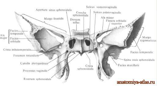
The front and part of the lower surface of the body face the nasal cavity. In the middle of the anterior surface of the body, a vertically running wedge-shaped ridge protrudes, Crista sphenoidalis. Its anterior edge is adjacent to the posterior edge of the perpendicular plate, lamina perpendicularis, ethmoid bone. The lower segment of the crest is pointed, extended downwards, and forms a wedge-shaped beak, rostrum sphenoidale, which is wedged between the opener wings, alae vomeris. On the sides of the ridge lies a thin curved plate - a wedge-shaped shell, concha sphenoidalis. This shell, forming the anterior and partly inferior walls of the sphenoid sinus, sinus sphenoidalis, has a small opening - the aperture of the sphenoid sinus, apertura sinus sphenoidalis. Outside of the aperture, there are small depressions that cover the cells of the posterior part of the labyrinth of the ethmoid bone. The outer edges of these recesses are partially connected to the orbital plate of the ethmoid bone, forming a sphenoid-ethmoid suture, sutura sphenoethmoidalis, and the lower ones - with the orbital process, processus orbitalis, palatine bone.
sphenoid sinus, sinus sphenoidalis, a steam cavity, performs most of the body of the sphenoid bone and belongs to the air-bearing paranasal sinuses. Both right and left sinuses are separated from one another by the septum of the sphenoid sinuses, which continues anteriorly into the sphenoid crest. As in the frontal sinuses, the septum sometimes lies asymmetrically, as a result of which the size of both sinuses may not be the same. Through the aperture, the cavity of each sphenoid sinus opens into the nasal cavity. The cavity of the sphenoid sinus is lined with a mucous membrane.
small wings, alae minores, sphenoid bone with two roots depart in both directions from the anterior-upper corners of the body in the form of two horizontally located plates, at the base of which there is a rounded hole. It represents the beginning of the bone canal up to 5-6 mm long - the visual canal, canalis opticus. It contains the optic nerve n. opticus, and the ophthalmic artery, a. ophthalmica. Small wings have an upper surface facing the cranial cavity, and a lower surface directed into the cavity of the orbit and closing the upper orbital fissure from above, fissura orbitalis superior. The anterior margin of the lesser wing, thickened and serrated, is connected to the orbital part of the frontal bone. The posterior concave and smooth edge protrudes freely into the cranial cavity and is the boundary between the anterior and middle cranial fossae, fossae cranii anterior et media. Medially, the posterior edge ends with a protruding, well-defined, anterior inclined process, processus clinoideus anterior(part of the dura mater is attached to it, forming the diaphragm of the Turkish saddle, diaphragma sellae).
Large wings of the sphenoid bone, alae majores, depart from the lateral surfaces of the body of the sphenoid bone and are oriented outwards. The large wing has five surfaces and three edges. superior cerebral surface, facies cerebralis, concave and turned into the cranial cavity. It forms the anterior part of the middle cranial fossa and bears sulcular depressions, cerebral eminences and arterial sulci, sulci arteriosi(imprints of the relief of the adjacent surface of the brain and middle meningeal arteries). There are three holes at the base of the large wing: a round hole is located inward and anteriorly, foramen rotundum(the maxillary nerve exits through it, n. maxillaris). Outside and behind the round is an oval hole, foramen ovale (it passes the mandibular nerve, n. mandibularis, and the vasculature of the foramen ovale). Still outside and posterior to the foramen ovale is the spinous foramen, foramen spinosum(through it pass the middle meningeal artery, vein and nerve). Antero-superior, orbital surface, facies orbitalis, smooth, diamond-shaped, turned into the cavity of the orbit, where it forms most of its outer wall. The lower edge of this surface is separated from the posterior edge of the orbital surface of the body of the upper jaw; here the inferior orbital fissure is formed, fissura orbitalis inferior. Anterior, maxillary surface, facies maxillaris, a small area of triangular shape, bounded above by the orbital surface, and from the side and below by the root of the pterygoid process of the sphenoid bone. It is part of the posterior wall of the pterygopalatine fossa, fossa pterygopalatina. There is a round hole on the surface. Upper lateral, temporal surface, facies temporalis, somewhat concave, takes part in the formation of the wall of the temporal fossa, fossa temporalis(the temporalis muscle is attached to it, m. temporalis). From below, this surface is bounded by the infratemporal crest, crista infratemporalis, below which the surface is located, where the oval hole opens, foramen ovale, and a spinous foramen. It forms the superior wall of the infratemporal fossa fossa infratemporalis. Here begins part of the lateral pterygoid muscle, m. pterygoideus lateralis. The upper, frontal, edge is widely serrated, connects with the orbital part of the frontal bone in the sphenoid-frontal suture ( sutura sphenofrontalis). The outer sections of the frontal edge end with a sharp parietal edge, margo parietalis, which with the wedge-shaped angle of the parietal bone forms a wedge-parietal suture ( sutura sphenoparietalis). The internal sections of the frontal margin pass into a thin free margin, which is separated from the lower surface of the lesser wing, limiting the upper orbital fissure from below fissura orbitalis superior. Anterior, zygomatic edge, margo zygomaticus, serrated, connects with the frontal process, processus frontalis, zygomatic bone, forming a wedge-zygomatic suture ( sutura sphenozygomatica). Back, scaly edge, margo squamosus, connects to the wedge-shaped edge, margo sphenoidalis, temporal bone in the sphenoid-squamous suture ( sutura sphenosquamosa). Posteriorly and outwards, the scaly edge ends with the spine of the sphenoid bone, spina ossis sphenoidalis. Here is the site of attachment of the sphenomandibular ligament, lig. sphenomandibular, and bundles of muscles that strain the palatine curtain, m. tensor veli palatini. Inward from the spine of the sphenoid bone, the posterior edge of the large wing lies in front of the petrous part, pars petrosa, temporal bone and limits the sphenoid-stony fissure, fissura sphenopetrosa, medially passing into a torn hole, foramen lacerum. This slot is made cartilage tissue, forming wedge-stony synchondrosis, synchondrosis sphenopetrosa.
pterygoid processes, processus pterygoidei, depart from the junction of the large wings with the body of the sphenoid bone and go down. The pterygoid processes are formed by two plates - lateral and medial. lateral plate, lamina lateralis processus pterygoidei, wider, but thinner and shorter than the inner one (the lateral pterygoid muscle begins from its outer surface, m. pterygoideus lateralis). medial plate, lamina medialis processus pterygoidei, narrower, thicker and slightly longer than the outer. Both plates grow together with their front edges and, diverging posteriorly, limit the pterygoid fossa, fossa pterygoidea(here begins the medial pterygoid muscle, m. pterygoideus medialis). In the lower sections, both plates do not fuse and limit the pterygoid notch, incisura pterygoidea filled with pyramidal process, processus pyramidalis, palatine bone. free end the inner plate ends with a pterygoid hook directed downward and outward, hamulus pterygoideus, on the outer surface of which there is a furrow of the pterygoid hook, sulcus hamuli pterygoidei(the tendon of the muscle straining the palatine curtain is thrown through it, m. tensor veli palatini). The posterior-upper edge of the inner plate at the base expands and forms an oblong navicular fossa, fossa scaphoidea(bunches of muscles begin in it, straining the palatine curtain, m. tensor veli palatini). Outside of the scaphoid fossa is a shallow furrow of the auditory tube, sulcus tubae audilivae, which laterally passes to the large wing and reaches the spine of the sphenoid bone (the cartilaginous part of the auditory tube is adjacent to this groove). Above the scaphoid fossa and medial from it there is an opening leading to the pterygoid canal, canalis pterygoideus(vessels and nerves pass through it). The canal runs in the sagittal direction in the thickness of the base of the pterygoid process and opens on the maxillary surface of the greater wing of the sphenoid bone on the posterior wall of the pterygoid fossa. Under the outlet, along the anterior face of the pterygoid process, there is a pterygopalatine groove. The inner plate at its base gives off a flat, horizontally running vaginal process directed inwards, processus vaginalis, which is located under the body of the sphenoid bone, covering the wing of the vomer from the side. As a result of this, the groove of the vaginal process facing the wing is the vomerovaginal groove, sulcus vomerovaginalis, turns into the vomerovaginal canal, canalis vomerovaginalis. Outside of the process, there is sometimes a sagittally running small palatovaginal groove, Sulcus palatovaginalis. In the latter case, the sphenoid process of the palatine bone adjacent from below closes the groove into the canal of the same name (the nerve branches of the pterygopalatine ganglion pass through both canals, and the branches of the sphenoid-palatine artery also pass through the palatovaginal canal). Sometimes, from the posterior edge of the outer plate, the pterygoid process is directed towards the spine of the sphenoid bone. processus pterygospinosus, which can reach the specified awn and form a hole.