All people have bad and good days, happy and sad events, something happens that angers, offends, upsets or, on the contrary, leads to indescribable delight, causes fun and happiness. At such moments, our face is just a book in which we can read all our feelings.
But why does this happen? What is it about the structure of the face that allows us to be so different, lively, interesting and multifaceted in expressing emotions? It turns out that this is a merit different types muscles. We'll talk about them in this article.
The physiology of muscle work was studied by the following scientists of the 18th-20th centuries:
- Luigi Galvani - discovered the phenomenon of electrical impulses in muscles and animal tissues.
- Emile Dubois-Reymond - formulated a law reflecting the effect of current on excitable tissues
- N. E. Vvedensky - described and established the optimum and pessimum of electrical excitation in muscles
- G. Helmholtz, J. Liebig, Wislicenus, V. Ya. Danilevsky and others - studied and described everything in detail physiological characteristics functioning of muscle tissue, including heat transfer during physical activity and muscle nutrition.
At the present stage of development, almost all possible theoretical descriptions of any functional characteristics of muscle fibers have been formulated. Electrophysiology, biochemistry, anatomy and other sciences have contributed to the accumulation of an extensive knowledge base in this area, so important for medicine.
Number and definition of human muscles
In total, there are about 640 muscles in the human body, each of which performs its own special function. Muscle anatomy is a collection of complex structural parts.
Muscles (or muscles) are human organs, which are a set of (elongated cells) having a smooth or cross-striated pattern. They are held together by loose connective tissue. In the human body they form a whole system (striated tissues) and line many organs and vessels (smooth tissues).

Classification
According to the functions they perform, muscles are divided into the following groups:
- Abductors.
- Leading.
- Arch supports.
- Sphincters.
- Dilators.
- Rotators.
- Flexors.
- Extensors.
- Contrasting.
- Pronators.
There is also a classification of muscles according to their location in the human body. So, they distinguish:
- (superficial and deep);
- limb muscles;
- head muscles (facial and chewing muscles).
Form
Based on this feature, 7 main muscle groups are distinguished, with each group localized and functional in a specific part of the human body.
- Fusiform.
- Square.
- Flat.
- Direct.
- Triangular.
- Cirrus.
- Circular.
Muscle anatomy
Each muscle has approximately the same internal structure: the outside is covered with epimysium, a special sheath substance produced by connective tissue. From the inside, it is a set of muscle bundles of different orders, which are united by endomysium - connective tissue. In this case, a row approaches each muscle blood vessels and capillaries for sufficient oxygen supply during operation. Veins take away decay products and carbon dioxide. The nerves that penetrate the fibers provide conductivity, excitability and a quick and high-quality response (work).
The muscle cells themselves have several nuclei, since during active work capable of producing thermal energy due to numerous mitochondria. Muscles owe their ability to contract to special proteins: actin and myosin. They provide this function by causing contraction of the myofibril - the contractile part of the muscle fiber.
The most important functions of muscle fibers are contractility and excitability, provided by the joint interaction of nerves and protein structures and controlled by the central nervous system (brain and spinal cord).

Head muscles
This group includes several main types. The main ones are:
- facial muscles (facial muscles) - responsible for facial expressions and external manifestations of emotions;
- chewing - perform the same function.
In addition to them, muscles are distinguished:
- eyeball;
- auditory ossicles;
- language;
- sky;
- pharynx.
The peculiarity of the structure of all the muscles of the head, except for the cheek, is the absence of fascia - a special “bag” in which all the muscles are located and which is attached directly to the bones. Therefore, the vast majority of them are attached to the bones on one side, and freely flow directly into the skin on the other, tightly intertwined with it into a single structure.
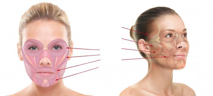
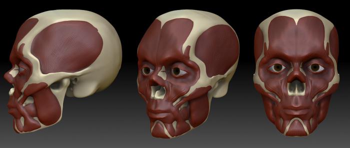
Mimic muscles of the face: types
The most interesting and clearly showing their work externally are the facial muscles. Due to the function they perform, that is, the ability to form human facial expressions, they got their name - facial muscles.
There are quite a lot of them. After all, you only have to remember how bizarre and varied the expressions of our emotions can be to understand that such work cannot be done alone or together. Therefore, facial muscles act in whole groups, and there are 4 of them in total:
- Forming the cranial vault.
- Forming the circumference of the mouth.
- Encircling the nose.
- Forming the circle of the eyes.
Let's look at each group in more detail.
Muscles of the cranial vault
The facial muscles of the head, which form the cranial vault, are represented by the occipitofrontal muscle, attached to the tendon helmet. The helmet itself is a tendon that conventionally divides the muscle into two parts: occipital and frontal. The main function performed by such facial muscles of the head is the formation of transverse folds of skin on the human forehead.
This group also includes the anterior and posterior auricular muscles. Their main action is to allow the auricle to move up, down, forward and backward.
The transverse nuchalis muscle is part of the structures of the cranial vault. The main function is the movement of the skin on the back of the head.
Muscles that form the circumference of the eye
These are the most expressive facial expressions. Their anatomy does not imply the presence of fascia, and the shape of such structures is different.
- The orbicularis muscle completely encircles the eyeball in a circle under the skin. Consists of three main parts: orbital, eyelid and lacrimal. Action - opening and closing eyes, controlling tear flow, lowering eyebrows, smoothing wrinkles on the forehead.
- The facial muscles that wrinkle the eyebrows are attached from the frontal bone to the skin of the eyebrows. Function: formation of longitudinal folds on the bridge of the nose.
- The proud muscle - the name itself speaks volumes - forms transverse folds at the base of the nose, giving the face an expression of pride and inaccessibility.
Such facial muscles allow people to express their emotions only with their gaze, eyes and skin around them. A lot can be said without words thanks to such structural features human body.

Muscles that form the circumference of the mouth
No less important are other facial muscles. The anatomy of this group of muscles is represented by a circular structure surrounding the mouth opening. There are several main muscles operating here, which are antagonistic to each other. This means that some of them expand the mouth opening, and some, on the contrary, narrow it.
- A muscle of the mouth called the orbicularis. Action: narrowing of the mouth and moving the lips forward.
- Zygomatic muscles (major and minor). Functions: Allow the corner of the mouth to move up, down and to the side.
- The peculiarities of the facial muscles of the mouth are that they allow it to move. For example, at the base of the upper jaw there is a muscle that allows you to lift upper lip. Nearby is one that raises the wing of the nose.
- Buccal muscle. Meaning: pulls the corner of the mouth to the side, with simultaneous contraction on both sides, allowing the inner surface of the cheeks to be drawn towards the jaw.
- Laughter muscle. Action: Allows the corners of the mouth to stretch laterally.
- Two mental muscles. The peculiarities of facial muscles of this type are that one of them is unstable and can be reduced. Function: provide movement of the skin of the chin, and also pull the lower lip forward.
- Muscle that lowers the lower lip. Meaning according to the name.
These are all the main oral facial muscles, the anatomy of which allows a person to smile, talk, express joy and dissatisfaction, and move the mouth.
Muscles surrounding the nose
This group includes only two main muscles:
- nasal muscle, consisting of an internal and external part. Action: provide movement of the nostrils and nose;
- muscle that pulls the nasal septum down.
Thus, there are only two facial muscles in the circumference of the nose. Their anatomy is no different from the others discussed above. In general, the listed muscle groups of the eye, mouth, nose and cranial vault are the main components of facial expressions. Thanks to the presence of these muscles, people are able to convey a range of feelings, communicate with each other even without words, and reinforce phrases with the necessary visual expression.
Facial muscles are very important structures, which are also responsible for the formation of wrinkles during the aging process. That is why all centers engaged in plastic surgery and similar procedures invite highly qualified specialists who know muscle anatomy well.
Chewing muscles: varieties
Facial and chewing muscles are the main components of the face and head. If the first group includes 17 different structures, then the second group includes only 4. However, it is these four masticatory muscles that play an important role in human life, as well as in maintaining a beautiful, young oval face. Let's look at exactly what structures belong to them.
- The chewing muscle is the strongest muscle trained by a person during food intake. It is located in two parts: deep and superficial. It starts from the zygomatic arch and attaches to the muscles of the lower jaw.
- Temporal - starts from the process temporal bone and extends to the lower jaw.
- Pterygoid lateral - consists of two parts: the upper and lower heads. It starts from the area of the sphenoid bone and ends in the muscles of the lower jaw, forming a complex interweaving with them.
- Pterygoid medial - also located from to the lower jaw.
All these muscles are united by the common functions they perform, which we will now consider.
Functions
Naturally, since the muscles belong to the group of chewing muscles, their action will be corresponding: ensuring versatile movement of the jaw:
- Chewing - the lower jaw is raised and pushed forward.
- Medial - provides lateral and other movements of the lower jaw.
- Lateral - has similar functions to the medial.
- The temporal lobe is the main assistant in chewing movements. Pulls back the lower jaw, which is pushed forward, and also allows it to rise up until it closes with the upper one.
In addition, it is the temporalis muscle that gives a person a tired, tired and haggard appearance. If you are in a state for a long time nervous tension, acute experiences and stress, the body will begin to lose weight, and the face will take on a corresponding haggard expression. This occurs due to the fact that the temporal muscle becomes thinner and, tightening the skin of the face, visually changes its relief.
Thus, we can conclude that the facial and masticatory muscles are the constructors of our face, allowing us to embed any expressions, make various movements and change various grimaces. They also allow chewing, which is undoubtedly one of the most important life processes of most living beings, including humans.
Among the ways of expressing feelings and emotions, facial expressions occupy one of the leading places. Facial muscles, changing the position of eyes, lips, eyebrows, reflect the internal state of a person. Mastery of facial expressions allows you not only to convey your internal state to your interlocutor, but also to emphasize the expressed thought and highlight the rhythm of the sound. The science of anatomy considers facial muscles as part of the human muscular system that performs important physiological functions. The branch of psychology that studies facial expressions allows you to read a person; the information carried by the muscles of a person’s face is almost impossible to fake. Even in the language of facial expressions there are obscene expressions, so to modern man If you are forced to communicate with a huge number of people every day, you should not only learn to read secret signs faces and gestures, but also to master the culture of non-verbal communication.
It is customary to begin acquaintance with non-verbal means of receiving and transmitting information from Facial muscles differ from skeletal muscles in that they do not have double attachment to the bones, they are necessarily woven into the skin at one or two ends, and they can also connect to the mucous membrane. Their contraction causes areas of the skin to move. Relaxation of facial muscles will lead to the return of the skin to its previous state, thanks to the elasticity of the skin. Antagonists have less effect on the work of facial muscles than on the functioning
At its beginning, visceral muscles were related to internal organs. In the process of development and a number of transformations, it degenerated into the skin muscles of the neck area, and subsequently, divided into bundles, into the muscles of the face.
Many muscle movements on a person’s face occur without conscious control over them. It takes extraordinary skill to keep a smile on your face when you are in a bad mood and to hide signals of sadness, much less to simulate emotions well.
It’s easier to understand the abundance of facial muscles if you divide them into groups. The frontalis muscle is a broad flat muscle, it is responsible for raising the eyebrows, its work leads to the formation of a series of frontal transverse folds.
The orbicularis oculi muscle belongs to the circular muscles, surrounding the eye, it controls the eyelids, looking sideways is also its merit.
The small muscle that wrinkles the eyebrows originates at the nasal septum, with its second end attached to the skin in the area of the middle of the eyebrow. Responsible for a frown and accompanies a sad mood. When the muscle lowers the tips of the eyebrows, a pair of vertical folds appears above the root of the nose, and the eyebrows converge toward the nose.
The quadrangular muscle, which controls the upper lip, has three sections, which are located on both sides of the nose. An expression of ridicule or contempt is produced when this muscle causes the upper lip to stretch.
There is a circular muscle around the mouth. It starts from the corners of the mouth from the small mouth and leads to bending and clenching of the lips.
The muscle, called the lower incisor, begins at the bottom of the cheek and is attached to the lower lip, it shades it down.
The chin muscle is located on the cheek. Her task is to pull her lower lip upward, protruding, giving her face a dissatisfied expression.
Responsible for lowering the lower lip single muscle. It starts at the lower edge of the jaw and ends at the corners of the mouth. It turns out to be a frowning grimace when the muscle pulls the corners of the mouth down.
The zygomatic arch has the zygomatic muscle, which stretches to the corners of the mouth. A pleasant smile appears on her face when she pulls up the corners of her mouth.
Facial muscles, constantly displaying certain emotional states a person, leave a pattern in the form of wrinkles on his skin. Psychologists involved in physiognomy have learned not only to accurately recognize a person’s character in the wrinkles of a person’s face, but even to read the history of its formation.
If you remember which muscles draw certain feelings on the face, then by the wrinkles around the eyes and mouth, on the forehead, cheeks and chin, by their direction and depth, you can judge what emotions most often possessed each person specific person. And then you can judge the character of the subject.
Unconsciously giving preference to certain feelings, a person not only colors his life and memories in a certain emotional color. Subsequently, everyone develops the habit of being in the most comfortable (familiar) state; this state does not always have a “+” sign. Don’t we know people who, constantly putting on the mask of “anger” or “irritation,” become so fused with it that it becomes part of their character? Facial muscles “remember” emotions, and if we forcibly try to restore one or another emotion on the face, then later it will take over ours to a greater or lesser extent. mental state. To concentrate, we wrinkle our foreheads, but to restore inner balance we start with transforming and controlling it.
LABORATORY WORK
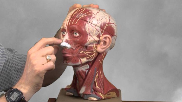
Muscles of the human body (practical work)Using drawings and anatomical descriptions, determine the locations (position of muscle groups and the movements they perform.
Rice. 35. Muscles of the head.
- frontal;
- temporal;
- circular eyes;
- circular mouth;
- chewing;
- sternocleidomastoid;
-occipital
Muscles of the head (according to Figure 35).
The facial muscles are attached to the bones, skin or only to the skin, the chewing muscles are attached to the bones of the fixed part of the skull and to the lower jaw.
Task 1. Determine the function of the temporal muscles. Place your hands on your temples and make chewing movements. The muscle tenses as it lifts the lower jaw upward. Find the masseter muscle. It is located near the jaw joints, about 1 cm in front of them. Determine: are the temporal and masticatory muscles synergists or antagonists?
Task 2. Get to know the function of facial muscles. Take a mirror and wrinkle your forehead, which is what we do when we are unhappy or when we are thoughtful. The supracranial muscle contracts. Find it in the picture. Observe the function of the orbicularis oculi and orbicularis oris muscles. The first one closes the eye, the second one closes the mouth.
27305150495 Sternocleidomastoid muscle on the anterior surface of the neck (according to Figure 35).
Task 3. Turn your head to the right and feel the left sternocleidomastoid muscle. Turn your head to the left and find the right one. These muscles turn the head left and right, acting as antagonists, but when contracted together, they become synergists and lower the head down.
Rice. 36. Muscles of the trunk and limbs:
A - front view. Muscles of the hand: 1 - flexors of the hand and fingers; 2 - biceps shoulder; 3 - deltoid muscle. Muscles of the trunk: 4 - pectoralis major; 5 - serratus muscle; 6 - abdominal muscles. Leg muscles: 7 - sartorius; 8 -- quadriceps femur; 9 - tibial muscles. B - rear view. Arm muscles: 10 - triceps brachii; 11 - extensors of the hand and fingers. Muscles of the trunk: 12 - trapezius; 13 - latissimus dorsi muscle; 14 - deep back extensors; 15 - gluteal. Leg muscles: 16 - biceps femoris; 17 - calf
III. Muscles of the torso in front (according to Figure 36).
Task 4. Find the big one pectoral muscle. This paired muscle tenses if you bend your arms at the elbow and forcefully fold them on your chest.
Task 5. Look at the picture of the abdominal muscles that form the abdominal press. They are involved in breathing, bending the body to the sides and forward, and transferring the body from a lying to a sitting position with fixed legs.
Task 6. Find the intercostal muscles: the external ones inhale, the internal ones exhale.
Muscles of the torso at the back (according to Figure 36).
Task 7. Find the trapezius muscle in the picture. If you squeeze your shoulder blades and throw your head back, it will be tense.
Fall 8. Find latissimus muscle backs. She lowers her shoulder down and puts her hands behind her back.
Fall 9. Along the spine are the deep back muscles. They straighten the body, tilting the body back. Determine their position.
Task 10. Find the gluteal muscles. They move the hip back. The deep back muscles and gluteal muscles in humans are most highly developed due to upright posture. They resist gravity.
27305345440Muscles of the arm (according to figures 28, 34 and 36).
Task 11. Find the deltoid muscle in the picture. She is above shoulder joint and moves the arm to the side to a horizontal position.
Task 12. Find the biceps and triceps brachii muscles. Are they antagonists or synergists?
Task 13. Muscles of the forearm. To understand their function, place your hand on a table, palm side down. Press it against the table, then clench your hand into a fist and unclench it. You will feel the muscles in your forearm contract. This happens because from the side of the palm on the forearm there are
h* muscles that flex the hand and fingers, and those that extend them are located on back side forearms.
Task 14. Feel near the wrist joint from the palmar surface of the tendons that go to the muscles of the fingers. Think about why these muscles are on the forearm and not on the hand.
Rice. 34. Flexor and extensor muscles:
- tendons of the head of the biceps muscle, shoulder;
- body of the biceps muscle;
- tail of the biceps muscle,
- radius bone;
- ulna;
- tail of the triceps brachii muscle; 7 - humerus; 8 - belly of the triceps muscle; 9 - blade; 10 - heads of the triceps brachii muscle
VI. Leg muscles (according to Figure 36).
Task 15. On the front surface of the thigh there is a very powerful quadriceps femoris muscle. Find it in the picture. She bends her leg hip joint and straightens the knee. To imagine its function, you need to imagine a football player hitting the ball. Its antagonist is the gluteal muscles. They move their leg back. Acting as synergists, both of these muscles hold the body in an upright position, fixing the hip joints.
There are three muscles on the back of the thigh that flex the leg at the knee.
Task 16. Rise up on your toes, you feel how tense you are calf muscles. They are located on the back of the lower leg. These muscles are well developed because they support the body in an upright position and are involved in walking, running, and jumping.
Attached files
Her job.
Movements in the joints. A muscle can pull up, but cannot push away bones, so different muscles perform opposite movements: some flex, others extend, some bring the arm to the body, others abduct, some rotate the bone clockwise, others counterclockwise. Muscles that act in the opposite direction are called antagonists muscles acting in one direction - synergists. It happens that the same muscle groups participate as antagonists in one movement, and as synergists in another.
Laboratory work
Muscles of the human body (practical work)
Using drawings and anatomical descriptions, identify the location of muscle groups and the movements they perform.
Rice. 35. Muscles of the head: 1 – frontal; 2 – temporal; 3 – circular eyes; 4 – circular mouth; 5 – chewing; 6 – sternocleidomastoid; 7 – occipital
I. Muscles of the head(according to Figure 35).
Mimic muscles are attached to bones, skin or just skin, chewable– to the bones of the fixed part of the skull and to the lower jaw.
Task 1. Define a function temporal muscles. Place your hands on your temples and make chewing movements. The muscle tenses as it lifts the lower jaw upward. Find the masseter muscle. It is located near the jaw joints, about 1 cm in front of them. Determine: are the temporal and masticatory muscles synergists or antagonists?
Task 2. Get to know the function of facial muscles. Take a mirror and wrinkle your forehead, which is what we do when we are unhappy or when we are thoughtful. Reduced supracranial muscle. Find it in the picture. Observe the function orbicularis oculi muscle And orbicularis oris muscle. The first one closes the eye, the second one closes the mouth.
II. Sternocleidomastoid muscle on the front surface of the neck (according to Figure 35).
Task 3. Turn your head to the right and feel the left sternocleidomastoid muscle. Turn your head to the left and find the right one. These muscles turn the head left and right, acting as antagonists, but when contracted together, they become synergists and lower the head down.
Rice. 36. Muscles of the trunk and limbs: A – front view. Muscles of the hand: 1 – flexors of the hand and fingers; 2 – biceps brachii; 3 – deltoid muscle. Muscles of the trunk: 4 – pectoralis major; 5 – serratus muscle; 6 – abdominal muscles. Leg muscles: 7 – sartorius; 8 – quadriceps femur; 9 – tibial muscles. B – rear view. Arm muscles: 10 – triceps brachii; 11 – extensors of the hand and fingers. Muscles of the trunk: 12 – trapezius; 13 – latissimus dorsi muscle; 14 – deep back extensors; 15 – gluteal. Leg muscles: 16 – biceps femoris; 17 – calf
III. Muscles of the trunk in front(according to Figure 36).
Task 4. Find pectoralis major muscle. This paired muscle tenses if you bend your arms at the elbow and forcefully fold them on your chest.
Task 5. Look at the picture of the abdominal muscles that form abdominal press They are involved in breathing, bending the body to the sides and forward, and transferring the body from a lying to a sitting position with fixed legs.
Task 6. Find intercostal muscles: external ones inhale, internal ones exhale.
IV. Muscles of the trunk from behind(according to Figure 36).
Task 7. Find in the picture trapezius muscle. If you squeeze your shoulder blades and throw your head back, it will be tense.
Task 8. Find latissimus dorsi muscle. She lowers her shoulder down and puts her hands behind her back.
Task 9. Along the spine are deep back muscles. They straighten the body, tilting the body back. Determine their position.
Task 10. Find gluteal muscles. They move the hip back. The deep muscles of the back and gluteal muscles in humans are most highly developed due to upright posture. They resist gravity.
V. Muscles of the arm(according to figures 28, 34 and 36).
Task 11. Find in the picture deltoid muscle. It is located above the shoulder joint and moves the arm to the side to a horizontal position.
Task 12. Find double-headed And triceps shoulder muscles. Are they antagonists or synergists?
Task 13. Muscles of the forearm. To understand their function, place your hand on a table, palm side down. Press it against the table, then clench your hand into a fist and unclench it. You will feel the muscles in your forearm contract. This happens because there are muscles located on the side of the palm of the forearm, flexing wrist And fingers, A straightening them are located on the back of the forearm.
Task 14. Feel near the wrist joint from the palmar surface of the tendons that go to the muscles of the fingers. Think about why these muscles are on the forearm and not on the hand.
VI. Leg muscles(according to Figure 36).
Task 15. On the front of the thigh