Deformation chest called a change in the shape of the musculoskeletal frame of the upper body. There are two main types of chest deformity in children: pectus excavatum and pectus carinatum.
What is associated with chest deformation in children, and what should parents do in case of such a diagnosis?
The health consequences associated with chest deformities in children depend on the type of deformity and its severity.
Funnel chest deformity in children is manifested in the retraction of the costal cartilages, resulting in the formation of a funnel, or depression, in the center of the chest.
There are 4 degrees of funnel chest deformity in children, depending on the depth of the funnel. With degree I deformation (a depression of no more than 2 cm), the child may not feel any symptoms of the disease at all. With higher degrees of deformation, the child may experience difficulty breathing, shortness of breath, and some disturbances in the functioning of internal organs due to their compression.
With keeled chest deformity in children, the sternum protrudes forward in the form of a keel, to which the ribs are attached at right angles. This deformation is often only a cosmetic defect. If the keeled deformity is severe, this can lead to problems in the functioning of the lungs, heart and other internal organs due to a violation of their relative position. In this case, it is necessary to conduct an examination and find out the features of the location and functioning of the child’s internal organs.
What can cause chest deformation in children?
Chest deformity in children is most often a congenital disease and is formed during the prenatal period, when the child is in the mother’s womb. Scientists have not yet found an exact answer why the baby’s chest is deformed. It is only known that the likelihood of this defect increasing when:
Negative heredity (the presence of this disease in the medical history of the child’s mother or father or their closest relatives);
exposure to teratogenic factors (negative factors affecting the pregnant woman and the fetus and causing disturbances in its development without affecting hereditary structures). These factors include the transfer expectant mother infectious diseases, taking antibiotics and other chemicals, contact with radiation, etc.
That is, expectant mothers need to follow standard recommendations: take care of themselves, do not contact sick people, use medications with caution, etc.
As for acquired chest deformation in children, it can be caused by serious illnesses suffered by the child (rickets, scoliosis, pulmonary diseases, etc.) and injuries to the upper part of the body.
How is chest deformity corrected in children?
Mild chest deformities in children are treated conservatively, without surgery. It consists of carrying out physiotherapeutic procedures, massage, therapeutic exercises and, if necessary, wearing the child with special compression devices, orthoses and dynamic compression systems.
In more serious cases, children are prescribed surgery to correct the shape of the chest. Previously, it was believed that the younger the child being operated on, the better, since the ability of children's tissue to regenerate is much higher than that of a teenager or adult. Therefore, operations to correct the shape of the chest were performed on children as early as preschool age. However, now most doctors agree that early surgical correction of the chest shape can lead to abnormal growth of the ribs, recurrence of the disease and the need for repeated surgery. Therefore, surgeons recommend performing the operation no earlier than 10-12 years for boys and 12-13 years for girls.
Breathing exercises and physical therapy for chest deformation in children
The first thing you need to do if you notice a chest deformity in a child is to consult a doctor (orthopedic surgeon or more specialized specialist). If a specialist confirms that the defect does not pose a risk to the child’s health, parents can fight the child’s chest deformation on their own, namely, work with the child breathing exercises and physical therapy. These methods cannot completely correct the defect, but can slow down its development.
Breathing exercises for chest deformation in children help correct the shape of the musculoskeletal frame, and also normalize the functioning of the heart and lungs. Before doing breathing exercises with your child, you should check with your doctor to see if there are any contraindications to these exercises.
Breathing exercises
1. Holding your breath. Stand straight, feet shoulder-width apart. Take a deep breath and hold your breath for as long as possible. Then exhale sharply through your mouth. Repeat 5-10 times.
2. Upper breathing. Can be performed both standing and sitting. Inhale slowly and deeply, making sure that your stomach remains still and your chest rises. Exhale sharply through your mouth, repeat 5-10 times.
3. Expansion of the chest. Stand up straight, take a deep breath, clench your fists and extend your arms in front of you at shoulder level. With a quick movement, move your arms back and smoothly return to the starting position. Repeat several times and exhale sharply through your mouth. During the exercise, the arm muscles should be very tense.
In addition to breathing exercises, it is very useful for children with chest deformities to perform exercises for development pectoral muscles: push-ups, pull-ups, exercises with dumbbells and elastic gymnastic tape. Strong muscles The chest will help slow down the deformation and even stop it; in addition, a developed muscular frame will visually correct the cosmetic defect and close the deformed chest.
Children with deformed chests benefit greatly from swimming; this sport helps develop the pectoral muscles and lungs and has very few contraindications. Volleyball, basketball and rowing are also often recommended for this disease, especially if the child shows interest in them.
Mild chest deformation in children usually does not affect their health, especially if parents take measures to correct the defect: they do breathing exercises with the child, teach him to play sports. And even if the degree of deformation is high, medicine suggests effective ways complete elimination of the defect, ranging from high-tech compression devices to modern operations with minimal intervention.
Grow up healthy!
Physical therapy exercises in the presence of pectus excavatum are, in fact, auxiliary in nature - the only effective way to correct this defect is surgical intervention. Gymnastics can only help in cases of mild anomalies that are not progressive in nature, as well as in situations where deformities are caused by external mechanical influence.
A set of exercises prescribed to patients may include walking around the hall for a couple of minutes without additional movements, and then raising your arms up at the height of inhalation and then lowering them down as you exhale (1-2 minutes).
From the main stance (arms down, legs together), the left leg is taken back, the arms are raised up and inhaled, then they return to the original position and exhale air from the lungs. After which they repeat everything too, but with the right foot. Do this 6-8 times with each leg. The pace is slow, the patient looks forward. From the same position, bend forward, spread your arms to the sides and inhale, returning to the starting position, exhale (also 6-8 times).
Sitting on the floor, legs apart and resting on your hands from behind, lift your pelvis off the floor, bend slightly, throwing your head back, and take a breath. Then they return to their original position and exhale the air collected in their lungs. Perform 4-6 times.
Lying on your back, placing your arms along your body, breathe deeply through your chest (repeat 3-4 times). In the same position, bend the feet alternately 8-10 times. After this, they spread their arms to the sides, bend the right leg, press the knee to the stomach, straighten the leg up, bend it again and lower it down. Then they do the same with the other leg. And so 8-10 times with each leg.
Without changing the starting position, bend your arms to your shoulders, spread your legs to the sides and inhale. Return to the starting position and exhale (also 8-10 times). Next, they perform the so-called “bicycle” exercise, that is, they seem to spin imaginary bicycle pedals in the air (8-10 times). Then they raise their arms up, while simultaneously bending their knees and pressing them to the stomach, and take a deep breath, return to the starting position and exhale (6-8 times).
The next position is lying on your stomach, arms along the body, leading forward through the sides, legs apart and inhaling. Return to the starting position and exhale (8-10 times).
Again, while on your stomach, spread your arms to the sides and make circular rotations back with them (repeat 8-10 times). Maintaining the original position on the stomach, put the hands with the gymnastic stick forward, place the stick behind the back, namely, on the shoulder blades, and inhale, and when returning to the starting position, exhale. The range of movements here is maximum. Perform them from 2 to 10 times.
This is the kind of exercise therapy complex that should be done: Example of an exercise scheme: 1. IP (starting position) – standing, legs apart, gymnastic stick below, grip at the ends. Twist: move the stick back behind your back with straight arms. Screw: move the stick forward, repeat 15-20 times, strain your abdominal muscles. 2.IP – basic stance, the stick stands vertically on the floor in front of you – inhale. Sit down, hang, leaning on the stick with both hands, stretching the back muscles - exhale. Return to IP. Repeat 8-10 times, the pace is slow. 3.IP – standing, legs apart, arms to the sides. With short springy movements, move your straight arms back, stretching the chest muscles and connecting the shoulder blades. Repeat 20-40 times, straining the abdominal and buttock muscles. 4.IP - lying on your stomach, arms to the sides. While inhaling, bend over, raise your head, shoulders and arms not high from the floor, trying to connect your shoulder blades, hold the position for 3 to 8 seconds. Return to IP – exhale. Repeat 3-5 times. Avoid excessive extension of the head back. 5.IP - the same, hands forward. As you inhale, bend over, bend your arms, pull your elbows back, connect your shoulder blades and hands to your shoulders. Hold the position for 3-5 seconds, return to IP – exhale. Repeat 6-8 times. 6.IP – bend your right leg at the knee, press your heel to your buttock with your right hand, stretching the muscles of the front thigh, 30-60 sec. Then do the same with the other leg. Then both feet together. 7.IP – the same. Hands under the chin. While inhaling, bend over and perform 8 movements with your arms, as when swimming breaststroke. Return to IP – exhale. Repeat 3-5 times. 8.IP – the same. Bend your legs, grasping your ankle joints with your hands. Keep your knees together, pressed to the floor. As you inhale, bend over and, straightening your legs, knee joints , stretch with straight arms behind your legs, connecting your shoulder blades and straightening your shoulders. Hold the position for 5-8 seconds. Return to IP – exhale. Repeat 3-5 times. 9.IP – the same, hands forward. As you inhale, raise your right straight arm low (to your ear), tensing the muscles of your upper back. Return to IP – exhale. Then with your left hand. Repeat 8 – 10 times with each hand. Raise both straight arms at the same time, without raising your head. Repeat 8-10 times. 10.IP – the same, hands “locked” behind your back. As you inhale, bend over. With springy movements, stretch your arms back, straightening your shoulders and bringing your shoulder blades together. Return to IP – exhale. Repeat 6-8 times for 3-5 approaches. 11.IP – the same. Hands along the body. As you inhale, bend over, connect your shoulder blades, and raise your arms and legs straight. Hold the position for 5-8 seconds to 1-3 minutes, do not hold your breath. For physically fit children, the exercise lying on their stomach can be performed with weights. (For example: dumbbells weighing 0.5 kg). 12.IP – lying on your back. As you inhale, raise your straight legs to a vertical position. As you exhale, lower your legs behind your head, stretching your back muscles. Return to IP – inhale. Repeat 4-6 times. 13.IP the same. As you exhale, raise your head, straining your abdominal muscles and lower your lower back as far as possible to the floor, pull your toes towards you, hold the position for 2-3 seconds. Return to IP – inhale. Repeat 8-10 times 14.IP the same. Legs are bent at the knees. Keeping your shoulders and head still, pull your legs towards your chest. Repeat 10-20 times, 2-3 approaches, fast pace. 15.IP – lying on your back, arms up. With a wave of your arms - sit down, raise your arms up, straighten up. Slowly return to the IP. Breathing is voluntary. Repeat 10 times. 16.IP – the same, legs pulled up vertically, lower back pressed to the floor. Raise your head and shoulders and with springy movements reach your hands towards your feet, tensing your abdominal muscles. Repeat 10 times, 2-3 approaches. 17.IP – the same, hands joined at the back of the head, legs bent at the knees, feet on the floor. As you exhale, raise your head with your hands, connect your elbows and, tensing your abdominal muscles, bring your elbows and knees as close as possible to each other. Hold for 1-2 seconds. Return to IP – inhale. Repeat 6-10 times. 18.IP – the same, arms along the body. "Soldier". Raise your head slightly (about 30 cm), arms, straight legs, pull your toes toward you, hold the tension for 30 seconds to 3 minutes. 19.IP – sitting on your heels, arms extended forward. The head and hands are pressed to the floor. The methodologist secures the child by the pelvis. Raise your straight arms and head not too high. Hold the position for 6-8 seconds. Don't throw your head back! Repeat 3-5 times. 20.IP – the same, hands are joined in a lock behind the back. Raise your straight arms as high as possible, hold the maximum position for 10-20 seconds, return to IP. Repeat 2-3 times. 21.IP – standing in the doorway. Arms to the sides, abdominal muscles tense. As you inhale, stretch your chest. Hold the position for 5-10 seconds, repeat 3-5 times. Perform the same exercise with your arms up. 22.IP – lying on your back, roller under the shoulder blades, relax for 10-20 minutes.
Such a serious disease, such as chest deformation, implies a rather serious change in its shape, which can be acquired or congenital. This pathology can cause serious disruption of the functioning of almost all organs of the chest, that is, the cardiac, respiratory and vascular systems. It should be noted that chest deformities develop in approximately 2% of children. While the child is still too small, such pathology is not noticeable. However, when the child reaches 3 years of age, this deviation becomes most obvious.
In medicine, there are certain classifications of chest deformities. For example, the appearance of such a severe pathology may be funnel-shaped. This form is the most common. In most cases it is characterized by a sunken and slightly depressed chest. Boys are susceptible to developing this disorder, since many children of primary school age have health problems due to the development of pulmonary emphysema. In a more severe form of the disease, a decrease or increase in pressure, a complex curvature of the spinal column, as well as a very serious disruption in the functioning of such important organs as the lungs and heart may occur.
Using the ICD 10 code, which is the officially accepted international classification of diseases, anyone can find out that there is a so-called keeled form of deformity, in which the chest protrudes quite strongly forward. Gradually, this pathology can manifest itself much more strongly, but it does not have a detrimental effect on the internal organs and spine. However, with this disease, the heart can take on a teardrop shape, and the patient experiences fatigue, shortness of breath and rapid heartbeat.
Considering the types of chest deformities that exist today, it is worth highlighting the flat shape. This deviation is mostly characteristic of children who have a so-called asthenic physique (long limbs, tall stature and narrow shoulders). It is this form of deformation that causes the presence of frequent colds and developmental delays in the child.
In accordance with the international classification of diseases existing today, the presence of deformation is distinguished, which is a partial or complete cleft of the chest. However, this pathology is extremely rare. A rather severe congenital condition gradually worsens over time. Its main danger is that the front part of the heart is not protected by the ribs, but is located under the skin. Because of this, you can even view the beat of this organ. It is this form of the disease that requires surgery.
Quite rare is a curved pathology, which in medicine is most often called Currarino-Silverman syndrome. And probably the rarest is the so-called costomuscular defect, which is called Poland syndrome.
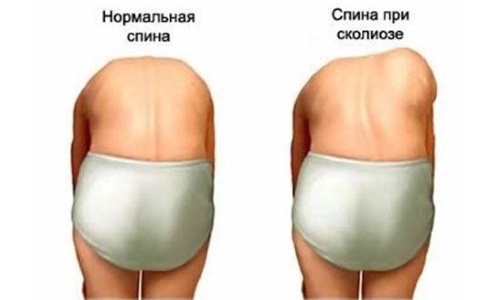
This severe pathology is characterized not only by serious deformation of the chest, but also by the presence of significant changes in the spinal column, internal organs and muscles. With the development of this pathology, the ribs and spine may become displaced. More inconvenience is caused by the so-called asymmetrical plano-concave deformation, which can be posterior, anterior or lateral.
Main causes
Among the most common causes of the development of such a serious illness are acquired pathologies and birth defects. This may be the presence of an emphysematous chest, which develops against the background of the appearance of pulmonary emphysema. In addition, deformation may appear due to previously suffered diseases of the pleura, which are chronic. However, most common cause The appearance of such a serious pathology is due to genetic factors that are gradually laid down precisely at the time when the baby is in the womb. In certain cases, the development of such a pathology is influenced by such common ailments as, for example, tuberculosis, rickets, scoliosis, various injuries and certain lung diseases.
This disease causes a significant displacement of all organs located in the chest cavity, which leads to too rapid heartbeat and difficulty breathing. Quite often the symptoms of the pathology are expressed by vegetative disorders, weakened immunity, as well as a fairly significant lag in physical development.
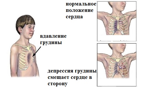
During the development of the disease, the patient may experience dysfunction of the respiratory, vascular and cardiac systems, scoliosis, and kyphosis. Due to the retraction of the sternum, asymmetrical posture may appear, which is a sign of the development of lateral curvature of the spinal column. In addition, during the study, a physician can identify a pathological process occurring directly in the stomach area. If the chest cavity increases, the diaphragmatic openings gradually weaken, which in the future leads to the development of a hernia.
To correctly diagnose the disease, specialists use MRI. This procedure gives a 100% diagnostic picture. Instrumental diagnosis of the disease may include radiography, which helps to identify the degree of development of the pathology. With more modern CT scans, doctors can examine mediastinal shifts, bone defects, and view the degree of compression of the lungs.
Magnetic resonance imaging allows you to obtain the most extensive information about all pathological processes that occur in bone and soft tissues. If there is a suspicion of dysfunction of the heart and lungs, the attending physician may recommend X-rays of the organs, echocardiography, or cardiac monitoring using the Holter method.
Chest deformation can be treated correctly and promptly using a variety of methods. In this case, everything depends on its form, degree of development, and also on whether there are disorders in the respiratory, vascular and cardiac systems.
At home, you can quite successfully use the most modern pharmaceutical treatment methods. However, such treatment does not provide sufficient correction large quantity violations. Medicines are used only to remove as quickly as possible many painful symptoms of a severe pathology. If the deformations are not too large, this suggests that the disease is only at the first stage. In this case, long-known and effective conservative methods are used.

Proper treatment of chest deformity at home involves the use of special exercises. In addition, the doctor may recommend massage, physiotherapeutic procedures, swimming, and wearing a special corset.
If a doctor recommended a patient to perform a procedure such as physical therapy, then only he should select the most effective exercises which will help to gradually eliminate pain. When performing a massage, it is recommended to follow certain rules. For example, before starting medical procedure The patient should definitely do some relaxation exercises or take a warm bath.
The massage itself is performed directly in pathological areas. It is in the place of deformation where the compaction has appeared that the massage therapist performs light and stroking movements, and in the areas of the existing bulge he presses harder with his hands. All massage exercises can be used only after agreement with the attending physician who diagnosed the patient’s condition. Massage is performed only by a qualified specialist.
In the presence of the so-called funnel-shaped pathology, which is most often congenital, the most effective vacuum bell method is used. During its application, a vacuum is created directly above the funnel, which helps to gradually pull it out. If this method does not give the expected result, specialists may prescribe the patient a procedure such as, for example, sternochondroplasty. It is used to treat diseases in children aged 6 - 7 years. In this procedure, the surgeon inserts a plate by excising the costal cartilage and making small incisions in the chest. In any case, after performing this manipulation, scars remain on the patient’s body, but this method is the most effective, and the baby’s parents will not need to think about how to correct the chest defect.
In the presence of deformity that is at stages 2 and 3 of development, only time-tested surgical treatment methods are used. The most popular in this case is the so-called minimally invasive surgical intervention using the Nuss method. Before performing it, the doctor must tell you how to relieve pain so that the operation is successful. The main advantage of this procedure is that after it there are no scars on the patient’s body. The essence of this method is to make very small cuts. Then specially made metal plates are inserted into them to help straighten the deformed chest. These plates are placed for approximately 4 years until the sternum takes its natural shape.
Prevention measures
To protect the chest from deformation, you need to try to save it from burns and severe injuries. It is very important to get rid of various chronic lung diseases in a timely manner.
For example, at school age, a child should strengthen his spinal muscles and do a full range of exercises every day. simple exercises physical therapy, pumping up the abs and playing sports. This way, you can keep your muscles toned, which will help protect your chest from curvature. It should be remembered that thoracic deformity is a very dangerous disease, the presence of which in most cases leads to quite serious disruptions in the functioning of internal organs.
In case of developmental defects (so-called malformations), which is pectus excavatum, the only means of correcting the defect is surgical intervention.
Therapeutic exercise, massage and sports They help only with deformations resulting from external mechanical influence.
Vitamin and hormonal preparations can help if the deformity is caused by metabolic disorders or general hormonal development
However, this does not mean that exercise is excluded as an ineffective method. Moreover, sports, massage, and gymnastics are a necessary (but not sufficient!) attribute of treatment; they are necessary for the patient’s complete recovery.
The use of the Nuss method in the correction of pectus excavatum allowed the patient to engage in sports during the recovery period, which made the correction of deformities even more effective. Therapeutic exercise and massage are indicated during the period when the plate is installed on the patient: it promotes muscle development as well as the formation correct posture. Sufficient attention to physical exercises allows you to achieve excellent results in the correction of pectus excavatum.
Exercises for chest deformities
According to statistics, for every 1000 children, 6-8 have congenital chest deformities. The cause of chest deformities is considered to be an abnormal growth of cartilage between the ribs and the sternum, which bends the sternum inward or outward. In the first case, the anomaly is called a funnel-shaped deformity (Pectus Excavatum), in the second - a keeled deformity (Pectus Carinatum). In some cases, these deformities can be effectively treated using non-invasive methods, including physical therapy.
As the muscles are trained, the chest deformity can be reduced, or at least not worsened. In addition, intensive pumping, especially in the torso area, allows you to increase muscle mass and create a good cosmetic effect - the curvature becomes less noticeable.
All physical exercises are aimed at solving the following problems:
- Increase mobility and flexibility of the spine and chest.
- Stretch all shortened structures.
- Strengthen the muscles for a more amplitude movement of the chest.
- Restore normal posture.
Exercises mobilize joint mobility, stretch soft fabrics around the sternum so that they do not interfere with the movement of the chest. In addition to the mobility of the joint-ligamentous apparatus, muscle tone improves.
Suggested Exercise Program
Stretching and mobilization exercises
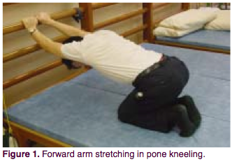
The patient is positioned on bent knees, arms extended forward. Holding the wall bars at a height of 50-80 cm, slowly lower top part body, and move your shoulder blades towards the floor. You should feel a stretch in the front armpit and shoulder. Hold for 8 seconds (you can breathe deeply, increasing the chest stretch), then relax. Repeat 20 times. Such approaches should be done 4 times a day.
The purpose of the exercise is to stretch all the anterior muscles of the chest, especially the pectoralis major muscle.
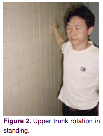
The patient stands sideways to the wall. Place the hand closest to the wall on the wall at a level slightly above the shoulder. Rotate your pelvis in the opposite direction, without lifting your hand from the wall. The stretch should be felt in the front of the shoulder and upper chest. Hold for 8 seconds. Then you can relax and return to the starting position. Rest and continue with the other hand. You need to do 4 sets of 20 repetitions per day.
This exercise provides the greatest range of motion of the thoracic vertebrae, allowing you to stretch the ligaments, muscles and joints in the sternum area.
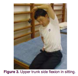
The patient is in a sitting position. Side bends are performed. When bending to each side, the opposite arm reaches overhead in the direction of the tilt. You should feel a stretch on the opposite side of your torso. Hold the position for 8 seconds (during this time you can breathe deeply), return to the starting position. Repeat exercises 20 times 4 times a day. The purpose of the exercise is the same as in the second exercise.
Strength exercises
4, 5. Exercise with weights in a lying position.
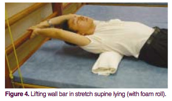
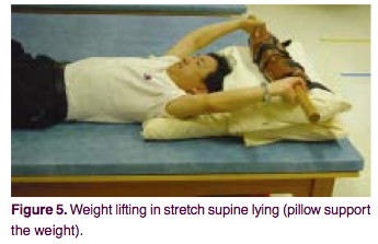
The patient is positioned in a supine position. You need to place a polyurethane foam roller about 10 cm in diameter under your back. If using a roller is too painful, just lie flat. Stretch your arms back behind your head and grab the bar of the wall bars or some heavy load at a distance of 10 cm from the ground. After this, take a deep breath and try to apply maximum effort, as if lifting a wall bar or a load. Hold the position for 8 seconds, then relax. 10 such repetitions constitute one series. You need to do 3 series 4 times a day.
Purpose of the exercise:
In this position, when the arms are fixed, the anterior chest wall rises mainly due to the pectoral muscle. Maximum tension when lifting force is applied causes the surrounding muscles involved in the breathing process to mobilize. A cushion placed under the back helps to expand and straighten the thoracic spine, correcting thoracic kyphosis.
6. Upper body stretch while lying on your stomach.
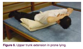
The patient lies on his stomach. You can place 1-2 pillows under your stomach. Hands behind your head. The legs can be fixed on the wall bars. Inhale deeply and raise your upper body, keeping your hands behind your head. Bend over. Hold this position for 8 seconds, then relax. 10 such repetitions constitute one series. You need to do 3 series 4 times a day.
The purpose of the exercise is to strengthen upper muscles back, balance muscle strength. This helps prevent thoracic kyphosis and maintain beautiful posture.
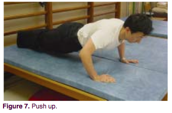
The patient lies on his stomach and begins to do push-ups. The easiest option is to lift only the torso, the more difficult is to lift the whole body, and the most advanced option is to clap your hands in the upper phase of the push-up, lifting your hands off the floor. Start with the first option. If it turns out easy, move on to the next one. One series is 10 repetitions. After each episode there is a rest. In total, you need to do 30 repetitions 4 times a day.
The purpose of the exercise is to generally strengthen the chest muscles. With intense loads, bone mineralization also improves; power pumping stimulates the formation of a beautiful silhouette.
8. Move your arms up and down along different sides of the body.
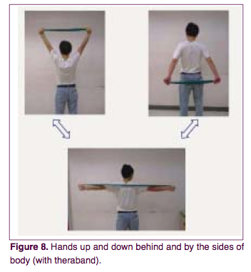
The patient is in a sitting or standing position. Arms extended. In your hands you need to hold the ends of an elastic band or any other elastic cord, its elasticity allowing you to do a maximum of 10 repetitions of the exercise, no more. Slowly move your arms back, lowering them to the level of your buttocks. Hold for 3 seconds. Then also slowly return your arms over the top to their original position. Repeat 10 times (1 series). After that, rest and do 2 more episodes. As a result, you need to do 30 repetitions 4 times a day.
The exercise serves to strengthen the neck, shoulders, upper back, and pectoral muscles. It can be considered as a stabilizing exercise for the upper chest.