The scapula is a flat bone. The scapula has three angles (upper (angulus superior), lower (angulus inferior) and lateral (angulus latera-lis)) and three edges (upper (margo superior), having a notch (incisura scapulae), lateral (margo late-ralis) and medial (margo medialis)).
There are concave (anterior costal (facies costalis)) and posterior (convex) surfaces (facies posterior). The costal surface forms the subscapular fossa. The posterior surface has the spine of the scapula (spina scapulae).
The collarbone (clavicula) is s-shaped. The clavicle has a body (corpus claviculae), thoracic (extremitas sternalis) and acromial (extremitas acromialis) ends. The upper surface of the clavicle is smooth, and on the lower surface there is a cone-shaped tubercle (tuberculum conoi-deum) and a trapezoidal line (linea trapezoidea).
The humerus (humerus) has a body (central part) and two ends. The upper end passes into the head (capet humeri), along the edge of which runs the anatomical neck (collum anatomikum). Behind the anatomical neck are the large (tuberculum majus) and small tubercles (tuberculum minus), from which the ridges of the same name extend (cristae tuberculi majoris et minoris).
Between head and body humerus The thinnest part of the bone is located - the surgical neck (collum chirurgicum).
On the lateral surface there is a deltoid tuberosity (tuberositas deltoidea), below which there is a groove of the radial nerve (sulkus nervi radialis). The distal end of the humerus ends in a cleft (condilus humeri), the medial part of which is represented by the block of the humerus (trochlea humeri), and the lateral part by the head of the condyle of the humerus (capitulum humeri).
The bones of the forearm include the ulna and radius.
The radius has a body and two ends. The proximal end passes into the head of the radius (caput radii), on which there is an articular fossa (fovea artikularis).
Ulna (ulna). At its proximal end there is a trochlear notch (incisura trochlea-ris), ending in two processes: the ulnar (olecranon) and the coronoid (processus coronoideus).
The hand (manus) consists of the bones of the wrist (ossa carpi), metacarpus (ossa metacarpi) and phalanges (phalanges) of the fingers. The wrist (carpus) consists of eight bones arranged in two rows.
The first row is formed by the pisiform (os pisiforme), triquetrum (os triquetrum), lunate (os lunatum) and scaphoid bones (os scaphoideum). The second row of bones consists of the hamate (os hamatum), capitate (os capitatum), trapezoid bones (os trapez-oideum) and trapezium bone (os trapezium).
There are five metacarpal bones. They are divided into a body (corpus metacarpale), a base (basis metacarpale) and a head (caput metacarpale). Phalanges of the fingers. All fingers, with the exception of the thumb, have three phalanges: proximal, middle and distal. The phalanx is divided into a body, a base and a head.
-
Structure belts upper limbs. The scapula is a flat bone.
Upper The surface of the clavicle is smooth, and on the bottom there is a cone-shaped tubercle (tuberculum conoi-deum) and a trapezoidal line (linea trapezoidea). -
Structure belts lower limbs. The pelvic bone (os coxae) consists of three bones fused together: the ilium, the pubis and
There are symmetrically located protrusions on the front and back of the ridge: upper anterior (spina ilia-ca anterior superior), lower anterior... -
Classification of joints belts upper limbs and their characteristics.
All bones of the skull except the joint temporal bone with the lower jaw, forming a joint. Structure joints belts lower limbs. -
Structure joints belts lower limbs. The sacroiliac joint (articulatio sacroiliaca) is formed by the auricular joints on top. Classification of joints belts upper limbs and their characteristics. -
Next question." Structure belts upper limbs. The scapula is a flat bone. The blade has three angles ( upper(angulus superio. -
Structure belts upper limbs.
Structure belts lower limbs. The pelvic bone (os coxae) consists of three bones fused together: the ilium, the pubis and the ischium... more details ". -
Structure belts lower limbs. The pelvic bone (os coxae) consists of three bones fused together: the ilium, the pubis, etc.
Body sphenoid bone has six surfaces: front, bottom, top, rear and two side. -
Structure joints belts lower limbs.
IN upper floor articulates the articular surface of the temporal bone with top the surface of the articular disc, and in the lower one - the head of the lower jaw with the lower surface of the articular disc. -
One of the proofs of the unity of origin of all vertebrates is the presence of bilateral symmetry, a general plan buildings spine, skull, limbs, belts limbs and all other systems. -
Nucleic acids. Structure, types and functions of RNA. An RNA molecule is a polymer whose monomers are nucleotides.
Muscles belts upper Certainly-ste set in motion top limb V shoulder joint, among them the most important delta muscle.
Similar pages found:10
The scapula is a flat bone. The scapula has three angles (upper (angulus superior), lower (angulus inferior) and lateral (angulus latera-lis)) and three edges (upper (margo superior), having a notch (incisura scapulae), lateral (margo late-ralis) and medial (margo medialis)).
There are concave (anterior costal (facies costalis)) and posterior (convex) surfaces (facies posterior). The costal surface forms the subscapular fossa. The posterior surface has the spine of the scapula (spina scapulae).
The collarbone (clavicula) is s-shaped. The clavicle has a body (corpus claviculae), thoracic (extremitas sternalis) and acromial (extremitas acromialis) ends. The upper surface of the clavicle is smooth, and on the lower surface there is a cone-shaped tubercle (tuberculum conoi-deum) and a trapezoidal line (linea trapezoidea).
The humerus (humerus) has a body (central part) and two ends. The upper end passes into the head (capet humeri), along the edge of which runs the anatomical neck (collum anatomikum). Behind the anatomical neck are the large (tuberculum majus) and small tubercles (tuberculum minus), from which the ridges of the same name extend (cristae tuberculi majoris et minoris).
Between the head and body of the humerus is the thinnest part of the bone - the surgical neck (collum chirurgicum).
On the lateral surface there is a deltoid tuberosity (tuberositas deltoidea), below which there is a groove of the radial nerve (sulkus nervi radialis). The distal end of the humerus ends in a cleft (condilus humeri), the medial part of which is represented by the block of the humerus (trochlea humeri), and the lateral part by the head of the condyle of the humerus (capitulum humeri).
The bones of the forearm include the ulna and radius.
The radius has a body and two ends. The proximal end passes into the head of the radius (caput radii), on which there is an articular fossa (fovea artikularis).
Ulna (ulna). At its proximal end there is a trochlear notch (incisura trochlea-ris), ending in two processes: the ulnar (olecranon) and the coronoid (processus coronoideus).
The hand (manus) consists of the bones of the wrist (ossa carpi), metacarpus (ossa metacarpi) and phalanges (phalanges) of the fingers. The wrist (carpus) consists of eight bones arranged in two rows.
The first row is formed by the pisiform (os pisiforme), triquetrum (os triquetrum), lunate (os lunatum) and scaphoid bones (os scaphoideum). The second row of bones consists of the hamate (os hamatum), capitate (os capitatum), trapezoid bones (os trapez-oideum) and trapezium bone (os trapezium).
There are five metacarpal bones. They are divided into a body (corpus metacarpale), a base (basis metacarpale) and a head (caput metacarpale). Phalanges of the fingers. All fingers, with the exception of the thumb, have three phalanges: proximal, middle and distal. The phalanx is divided into a body, a base and a head.
STRUCTURE OF THE SKELETON OF THE LIMB BELT
In domestic animals (ungulates and carnivores), only one dorsal bone of the girdle is preserved in the shoulder girdle - the scapula. The two ventral bones of the girdle - the clavicle and the coracoid bone - have undergone reduction in digitally and hoof-walking animals and can remain in the form of processes on the tubercle and acromion of the scapula.
All three bones are preserved on the pelvic girdle: the dorsal - ilium - os ilium and the ventral - ischium - os ischii (back) and pubic - os ilium (front) bones.
Shoulder girdle - girdle of the thoracic limb - cingulum membri thoracici.
The shoulder blade - scapula - is a large lamellar bone, attached to the body with the help of muscles (Fig. 53). Located in the area of the first ribs. Triangular shape. The wide base of the scapula is directed upward, and in ruminants and horses it is complemented by a wide scapular cartilage - cartilago scapulae. The caudal and cranial edges of the scapula go from the corresponding corners of the base down to the narrowed part of the scapula, on which the flat glenoid cavity is clearly visible - cavitas glenoidalis. Above the glenoid cavity there is a rather wide neck of the scapula - collum scapulae. On the cranial edge of the scapula, the tubercle of the scapula - tuber scapilae (supraarticular tubercle - tuberculum supraglenoidale) is located above the articular cavity; a powerful biceps shoulder The coracondial process is visible on the medial side of the tubercle.
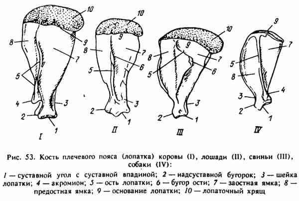
Rice. 53. Bone shoulder girdle(shoulder blade) of a cow (I), horse (I), pig (III), dog (IV)
The medial surface of the scapula is smooth, deepening towards the glenoid cavity - this is the subscapular fossa - fossa subscapularis. Above the subscapular fossa at the base of the scapula there is a serrated surface - fades serrata. Along the lateral surface of the scapula there is a spine of the scapula - spina scapulae; in its middle part it thickens, forming a tubercle of the spine - tuber spinae scapulae, which can be felt through the skin. The lateral surface of the scapula is divided by the spine of the scapula into the prespinous and infraspinous fossa - fossa supra-spinata and fossa infraspinata.
In ruminants, the spine of the scapula rises towards the glenoid cavity and breaks off sharply, not reaching the neck of the scapula, forming an acromion;
in horses, the spine of the scapula disappears towards the neck;
in pigs, the shoulder blade is in the form of an isosceles triangle, has a large lamellar on the spine triangular shape and a caudally directed tubercle of the spine. The spine of the scapula disappears at the neck;
in dogs, the spine of the scapula is high, with its elevated end hanging over the neck to the level of the glenoid cavity, forming a significant acromion.
Pelvic girdle - cingulum membri pelvini. In domestic animals, it fuses with the ventral bones, forming the pelvis - pervis. Each half of the pelvis is made up of an innominate bone - os coxae. The dorsal bone of the pelvic girdle - the iliac - os ilium - fuses with the ventral bones: in front lies the pubic bone - os pubis, behind - the ischium - os ischii. At the site of fusion of all three bones, a deep articular cavity is formed on each innominate bone - acetabulum, to which the free pelvic limb is attached. The ventral bones of the pelvis - the pubic and ischial bones - fuse with each other along the midline to form the pelvic fusion - symphisis pelvina - and form the floor of the pelvic cavity. The roof of the cavity is the sacrum and the first caudal vertebrae. The resulting pelvic cavity - cavum pelvis has an entrance formed from above by the sacral bone, on the sides - by the bodies of the iliac bones and below - by the pubic bones, and an exit, which is framed above by the first caudal vertebrae, and below by the ischial arch (Fig. 54).
The ilium has a columnar body - corpus ossis ilii, a lamellar wing - ala ossis ilii, which carries an articular (ear-shaped) surface for connection with the wing of the sacrum. The upper edge of the wing (cranial in ruminants and horses) has a lateral iliac tubercle (tuber coxae) and a medial sacral tubercle (tuber sacrale). From the base of the sacral tubercle along the body ilium to the ischial spine, which lies above the glenoid cavity, there is a large sciatic notch - incisura ischiadica major. From the auricular surface of the wing along the cranial edge of the body to the pubic bone there is a gentle iliac crest- crista iliaca - place of attachment of the muscles of the abdominal wall.
The pubic bone - os pubis - forms the cranial part of the pelvic floor, lies in front of the ischium. It has two parts: the caudal suture branch - ramus caudalis ossis pubis forms a pelvic fusion - symphisis pelvina - with the cognate branch along the midline and the cranial branch - ramus cranialis ossis pubis, which participates in the formation of the articular cavity of the pelvis. The cranial edge of the pubic bones forms the crest of the pubic bone - pecten ossis pubis, to which the muscles of the abdominal wall are attached.
The ischium - os ischii lies behind the caudal rami of the pubic bones, forming the caudal part of the pelvic floor and pelvic fusion. It has a body and a suture branch, which, together with the branches of the pubic bone, form a rather large closed opening - foramen obturatum. The posterior edge of the paired ischium forms the ischial arch - arcus ischiadicus. Lateral to it lie the ischial tuberosities - tubere ischiadica. They can be easily palpated and are used when measuring animals. From the lateral surface of these tubercles comes the lateral edge of the ischium, which is called the lesser ischiadica minor, it reaches the caudal edge of the ischium.
Features of the structure of the bones of the pelvic girdle.
The ruminant wings of the ilium with a well-defined macular and sacral tubercle are located in the frontal plane, slightly elevated along the cranial edge and overlap the wings of the sacral bone from above. The ischial tuberosities are very powerful with three eminences. The ischial spine and the obstructed foramen are significantly pronounced. The pelvic floor is concave, the ischial arch is deep.
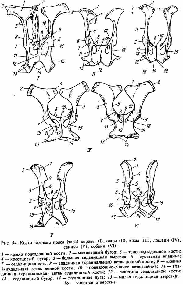
Rice. 54. Bones of the pelvic girdle (pelvis) of a cow (1), sheep (II), goat (III), horse (IV), pig (V), dog (VI)
In horses, the wings, like in ruminants, are located in a horizontal (frontal) plane with well-defined tubercles, overlying the wing of the sacrum. The ischial arch is flat, the ischial tuberosity, like the ischial spine, is small.
In pigs, the wings of the ilium are placed almost in the sagittal plane. They are elongated, adjacent to the wing of the sacrum on the lateral side. The ischial spine and ischial tuberosity are strongly pronounced, the ischial arch is deep.
In dogs, like in pigs, the wings are set in the sagittal plane, but the ischial spine is insignificant, the ischial arch is flat, with small ischial tuberosities. The pelvic floor is wide and flat. The marrow and sacral tubercle are poorly expressed.