Tubular bones They are long and short and perform the functions of support, protection and movement. Tubular bones have a body, a diaphysis, in the form of a bone tube, the cavity of which is filled in adults with yellow bone marrow. The ends of the tubular bones are called epiphyses. The cells of spongy tissue contain red bone marrow. Between the diaphysis and epiphyses are the metaphyses, which are areas of bone growth in length.
Spongy bones distinguish between long (ribs and sternum) and short (vertebrae, carpal bones, tarsus).
They are constructed of a spongy substance covered with a thin layer of compact. Spongy bones include sesamoid bones (patella, pisiform bone, sesamoid bones of the fingers and toes). They develop in muscle tendons and are auxiliary devices for their work.
Flat bones , forming the roof of the skull, built from two thin plates of a compact substance, between which there is a spongy substance, diploe, containing cavities for veins; the flat bones of the belts are built of spongy substance (scapula, pelvic bones). Flat bones serve as support and protection,
Mixed dice merge from several parts that have different functions, structure and development (bones of the base of the skull, collarbone).
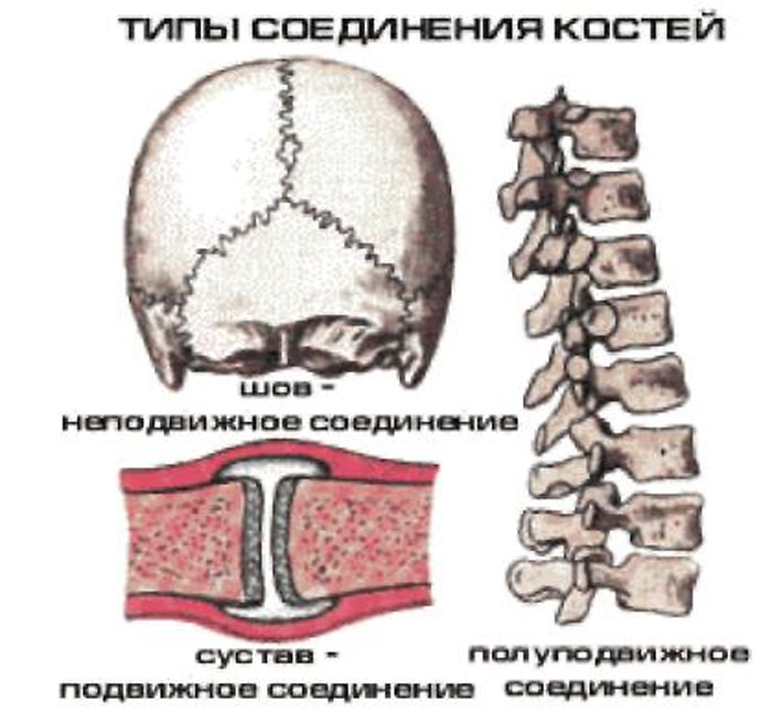
Question 2. Types of bone joints.
All bone connections can be divided into 2 groups:
continuous connections - synarthrosis (immobile or sedentary);
discontinuous joints - diarthrosis or joints (mobile according to function).
The transitional form of bone joints from continuous to discontinuous is characterized by the presence of a small gap, but the absence of an articular capsule, as a result of which this form is called a semi-joint or symphysis.
Continuous connections are synarthrosis.
There are 3 types of synarthrosis:
Syndesmosis is the joining of bones using ligaments (ligaments, membranes, sutures). Example: skull bones.
Synchondrosis is a connection of bones using cartilage tissue (temporary and permanent). The cartilage tissue located between the bones acts as a buffer, softening shocks and shocks. Example: vertebrae, first rib and vertebra.
Synostosis is the joining of bones through bone tissue. Example: pelvic bones.
Discontinuous joints, joints – diarthrosis . At least two are involved in the formation of joints articular surfaces , between which is formed cavity , closed joint capsule . Articular cartilage , covering the articular surfaces of the bones are smooth and elastic, which reduces friction and softens shocks. The articular surfaces correspond or do not correspond to each other. The articular surface of one bone is convex and is the articular head, and the surface of the other bone is correspondingly concave, forming the articular cavity.
The joint capsule is attached to the bones that form the joint. Hermetically closes the joint cavity. It consists of two membranes: outer fibrous and inner synovial. The latter secretes a clear liquid into the joint cavity - synovia, which moisturizes and lubricates the articular surfaces, reducing friction between them. In some joints, the synovial membrane forms, protruding into the joint cavity and containing a significant amount of fat.
Sometimes protrusions or inversions of the synovial membrane are formed - synovial bursae lying near the joint, at the junction of tendons or muscles. Synovial bursae contain synovial fluid and reduce friction between tendons and muscles during movement.
The articular cavity is a hermetically sealed slit-like space between the articular surfaces. Synovial fluid creates a pressure in the joint below atmospheric pressure, which prevents the divergence of the articular surfaces. In addition, synovia is involved in fluid exchange and strengthening of the joint.
Bone- a structural and functional unit of the skeleton and an independent organ. Each bone occupies a precise position in the body, has a certain shape and structure, and performs its characteristic function. All types of tissues take part in bone formation. Of course, the main place is occupied by bone tissue. Cartilage covers only the articular surfaces of the bone, the outside of the bone is covered with periosteum, and the bone marrow is located inside. Bone contains fatty tissue, blood and lymphatic vessels, and nerves. Bone tissue has high mechanical properties; its strength can be compared to the strength of metal. Relative bone density is about 2.0. Living bone contains 50% water, 12.5% organic protein substances (ossein and osseomucoid), 21.8% inorganic mineral substances (mainly calcium phosphate) and 15.7% fat.
In dried bone, 2/3 are inorganic substances, which determine the hardness of the bone, and 1/3 are organic substances, which determine its elasticity. The content of mineral (inorganic) substances in bone gradually increases with age, causing the bones of older and older people to become more fragile. For this reason, even minor injuries in old people are accompanied by bone fractures. The flexibility and elasticity of bones in children depend on the relatively higher content of organic substances in them.
Osteoporosis- a disease associated with damage (thinning) of bone tissue, leading to fractures and bone deformation. The reason is failure to absorb calcium.
The structural functional unit of bone is osteon. Typically, an osteon consists of 5-20 bone plates. Osteon diameter is 0.3 - 0.4 mm.
If the bone plates fit tightly to each other, then a dense (compact) bone substance is obtained. If the bone crossbars are loosely located, then spongy bone substance is formed, which contains red bone marrow.
The outside of the bone is covered with periosteum. It contains blood vessels and nerves.
Due to the periosteum, the bone grows in thickness. Due to the epiphyses, the bone grows in length.
Inside the bone there is a cavity filled with yellow bone marrow.
Classification of bones according to form:
- Tubular bones- have a general structural plan, they distinguish between a body (diaphysis) and two ends (epiphyses); cylindrical or triangular shape; length prevails over width; On the outside, the tubular bone is covered with a connective tissue layer (periosteum):
- long (femoral, shoulder);
- short (phalanxes of fingers).
- Spongy bones- formed predominantly by spongy tissue surrounded by a thin layer of solid matter; combine strength and compactness with limited mobility; The width of the spongy bones is approximately equal to their length:
- long (sternum);
- short (vertebrae, sacrum)
- sesamoid bones - located in the thickness of the tendons and usually lie on the surface of other bones (patella).
- Flat bones- formed by two well-developed compact outer plates, between which there is a spongy substance:
- skull bones (roof of the skull);
- flat (pelvic bone, shoulder blades, bones of the upper and lower limbs).
- Mixed dice- have a complex shape and consist of parts that differ in function, form and origin; due to their complex structure, mixed bones cannot be classified as other types of bones: tubular, spongy, flat (the thoracic vertebra has a body, an arch and processes; the bones of the base of the skull consist of a body and scales).
The bones that make up the skeleton make up approximately 18% of the total body mass.
Classification of bones is currently carried out not only on the basis of their structure, but also function and development. As a result, bones are distinguished between tubular, spongy, flat and mixed.
Tubular bones have the function of support, protection and movement. They are shaped like a tube with a medullary canal inside. The relatively thinner middle part of the tubular bones is called the body or diaphysis, and the thickened ends are called epiphyses. The thickening of the ends of long tubular bones is functionally justified. The epiphyses serve as the junction of bones with each other, and muscle attachment occurs here. The wider the surface of contact between the bones, the stronger it is; more stable connection. At the same time, the thickened epiphysis moves the muscle away from the long axis of the bone, as a result of which the latter approaches the attachment site at a large angle. This, according to the rule of parallelogram of forces, increases the force of the beneficial action of the muscle. Tubular bones are divided into long and short.
Long bones, the length of which significantly exceeds their other dimensions, constitute the proximal parts of the skeleton of both limbs.
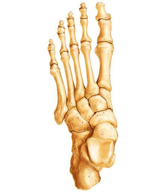
Short bones are located in the metacarpus, metatarsus, phalanges, etc. where greater strength and mobility of the skeleton are simultaneously required.
Spongy bones are divided into long, short, and sesamoid.
Long spongy bones (ribs, sternum) consist predominantly of spongy substance covered with a compact substance and serve the function of support and protection.
Short spongy bones (vertebrae, carpal bones, tarsals) consist mainly of spongy substance and serve as support.
Sesamoid bones (patella, pisiform bone, sesamoid bones of the fingers and toes) consist of spongy substance, develop in the thickness of the tendons, strengthen the latter and serve as a block through which they are thrown. This increases the shoulder for applying muscle force and creates more favorable conditions for its work. Sesamoid bones got their name from their resemblance to sesame seeds.
Flat bones form the walls of cavities containing internal organs. Such bones are curved on one side and convex on the other; their width and length significantly exceed their thickness. These are the pelvic bone, scapula, and bones of the skull.
Mixed bones lie at the base of the skull, have different shapes and development, the complexity of which corresponds to the variety of functions performed.
Among the flat and mixed bones of the skull, there are air-bearing ones, containing a cavity lined with mucous membrane and filled with air, which makes the bones lighter without compromising their strength.
The surface relief of the bone is not the same and is determined by the mechanical influence of neighboring organs. The vessels and nerves adjacent to the skeleton, muscles and their tendons leave marks on the bones in the form of grooves, notches, holes, roughnesses and channels. Areas on the surface of the bone that are free from attachment of muscles and ligaments, as well as articular surfaces covered with hyaline cartilage, are completely smooth. Surfaces of bones at places of attachment to them strong muscles elongated in the form of tubercles, tubercles and processes, increasing the area of attachment. Therefore, in people whose profession involves performing heavy physical activity, the surfaces of the bones are more uneven.
The bone, with the exception of the connecting surfaces, is covered with periosteum. This is a thin connective tissue membrane that is rich in nerves and vessels that penetrate from here into the bone through special openings.
Through the periosteum, the bone is nourished and innervated. The importance of the periosteum is to facilitate the attachment of muscles and ligaments that are woven into its outer layer, as well as to soften shocks. The inner layer of the periosteum contains bone-forming cells - osteoblasts, which ensure the growth of developing young bones in thickness.
When bones are fractured, osteoblasts form a callus that connects the ends of the broken bone, restoring its integrity.
Classification of compounds. The mobility of parts of the skeleton depends on the nature of the joints of the bones. The apparatus connecting the bones develops from the mesenchyme lying between the rudiments of these bones in the embryo. There are two main types of bone joints: continuous and discontinuous, or joints. The first are more ancient: they are found in all lower vertebrates and in the embryonic stages of higher ones. When bones are formed in the latter, their original material (connective tissue, cartilage) is preserved between them. With the help of this material, bones fusion occurs, i.e., continuous connection. At later ontogenetic stages, more advanced, discontinuous connections arise in terrestrial vertebrates. They develop due to the appearance of a gap in the original material preserved between the bones. Remnants of cartilage cover the articulating surfaces of bones. There is a third, intermediate type of joint - a semi-joint.
Continuous connections. A continuous connection - synarthrosis, or fusion - occurs when the bones are connected to each other by a continuous layer of tissue connecting them. Movements are limited or completely absent. Based on the nature of the connective tissue, connective tissue fusions, or syndesmoses, cartilaginous fusions, or synchondrosis, and fusions with bone tissue - synostoses, are distinguished.
There are three types of syndesmoses:
1) interosseous membranes, for example, between the bones of the forearm or lower leg;
2) ligaments connecting bones (but not connected to joints), for example, ligaments between the processes of the vertebrae or their arches;
3) sutures between the bones of the skull. Interosseous membranes and ligaments allow some displacement of the bones. At the sutures, the layer of connective tissue between the bones is insignificant and movement is impossible.
Synchondrosis is, for example, the connection of the first rib with the sternum through the costal cartilage, the elasticity of which allows some mobility of these bones.
Discontinuous joints - diarthrosis, articulation, or joint, is characterized by the presence of a small space (gap) between the ends of the connecting bones. There are simple joints formed by only two bones (for example, shoulder joint), complex, when the joint includes a larger number of bones (for example, the elbow joint), and combined, allowing movement only simultaneous with movement in other anatomically separate joints (for example, the proximal and distal radioulnar joints). The obligatory structural formations of the joint include the articular surfaces, the articular capsule, or capsule, and the articular cavity.
In addition to the obligatory ones, auxiliary formations may be found in the joint. These include articular ligaments and lips, intraarticular discs and menisci.
Skeleton, which is the axial organ musculoskeletal system, contains different types of bones. They differ in shape, structure and functions.
Features of the structure of bone tissue
This type of connective tissue has a typical structure. It consists of cells called osteocytes and a substance that fills the space between them. This fabric is unique in its own way chemical composition and properties. Organic substances, namely collagen fibers, give it elasticity. And mineral salts are strength. For example, the femur can withstand a load of several tons. And if inorganic substances are removed from bone tissue, it will easily crumble.
Types of human bones: classification signs
It is based on several signs. These are shape, size and structure. The types of bones also determine the function they will subsequently perform. The shapes are long, short and wide. The former contain a cavity inside filled with yellow bone marrow. This structure provides long strength and lightness. At their ends there is a spongy substance, between the elements of which there is red bone marrow. This is the basis of the body's hematopoietic cells. Short and completely formed by spongy substance.
Bones are also paired and have no analogues in the body. The first group mainly forms the skull. These include the temporal, zygomatic, parietal. Some bones of the belts and free limbs. These are the clavicles, shoulder blades, radial, humeral, pelvic. Examples of unpaired bones are the frontal, occipital, and mandibular.
Based on their location in the body, the bones of the skeleton and the torso are distinguished. The last group includes the spine, sternum and ribs. Also, according to this feature, the bones of the belts and free upper and lower extremities are distinguished. There are more than 200 of them in the human body.

Table: bone types
| Types of bones | Examples | Structural features |
| Long (tubular) | Femoral, tibial, humeral, radial, ulnar | The length of the bones of this species significantly exceeds the width. On top there is a connective tissue layer - the periosteum. Due to it, growth in thickness occurs. At the ends of the bone there is a spongy substance that contains red bone marrow. This is where blood cells are formed. The bone cavity is filled with yellow bone marrow. |
| Short | Frontal and parietal bones of the skull | The length and width of bones of this type are approximately the same. They are entirely formed by a spongy substance that covers a layer of compact substance. |
| Wide (flat) | Sternum, ribs, shoulder blades | The area of the bones exceeds the thickness. They are formed by two plates consisting of a compact substance, between which there is a spongy substance. Due to their large plane, they provide a basis for muscle attachment. |
What are mixed bones?
Very often, due to the complex structure of the bone, it cannot be classified as the main type. They are called mixed. These structures include the vertebrae, sacrum, and clavicle bones. They consist of several parts. Thus, the vertebra is formed by a body and processes, and the main function of this structure is to protect the spinal cord.

Types of bones and features of their connection
All bones in the human body are combined into a complex system using connections various types. The way they are attached to each other determines the function of the resulting structure. For example, the flat and wide bones of the skull are connected motionlessly. This method is called a seam. This connection allows you to create reliable protection for the brain. Different types of skeletal bones differ in their characteristics. For example, ulnar, radial and knee joint They move great. This movable connection provides the main function of this structure. It is to provide movement individual parts and the skeleton as a whole. The spine, which is the axial structure of the body, is a semi-movable joint. The whole point is that between him separate elements cartilaginous layers are located. This structure provides shock absorption when moving.
So, the main types of skeletal bones in the human body are tubular, short and wide. Their main differences lie in the features of the internal structure, shape, type of connection and function performed.