Under the control of fluoroscopy in direct and lateral projections, a needle with a plastic catheter put on it is inserted into the bile duct previously contrasted with PTCG. The needle is removed, a conductor is inserted through the catheter into the common bile duct or the distal intrahepatic duct.
External drainage of the biliary tract
After the catheter is removed, a drainage tube is inserted through the conductor, fixed to the skin and connected to the bile receiver. With obstruction of the biliary tract, especially in malignant tumors, one would expect that external drainage of the bile ducts in the preoperative period would lead to an improvement in the patient's condition and a decrease in the risk of developing renal failure after surgery. However, it is often accompanied by complications such as fluid and electrolyte loss, sepsis, and displacement of the drainage tube. As shown in a number of randomized controlled trials, short (1-2 weeks) drainage of the biliary tract before surgery for tumor obstruction of the biliary tract does not reduce mortality and does not improve the course postoperative period. Currently, the technique of percutaneous or endoscopic placement of endoprostheses, as well as the imposition of a biliodigestive anastomosis surgically, is sufficiently developed and there is practically no need for long-term drainage of the biliary tract with its physical and psychological side effects.
How to prepare for bile drainage?
Do not eat or drink for 4 hours before the procedure. This is because the procedure is done under sedation or general anesthesia - if your stomach is full, stomach contents can inadvertently pass into your lungs, which can be harmful. This can happen any time you have sedative medication or anesthesia, not just bile drains, and is the reason you are asked not to eat or drink.
If you are a diabetic, it is recommended that you consult with a radiology practice prior to fasting. You may need to stop taking medications that thin your blood as they increase your risk of bleeding. Examples include warfarin, clopidogrel, azasinthin, heparin, and enoxaparin sodium. If you think you may be taking any of these medications, please discuss this with your radiology practice prior to your procedure.
Internal and external drainage of the biliary tract
Bile duct catheterization allows a guidewire to be passed through the stricture into the distal common bile duct or into the intestine. A catheter (8-9 F) with holes above and below the constriction is placed in the area of the stricture. Bile enters the intestines or, with a catheter that opens outward, into a bile receptacle. Short-term (within a few days) external drainage of the biliary tract is used to prepare the patient for the installation of an endoprosthesis. The method can be used for the purpose of long-term decompression of the biliary tract, but the tube is left forever, even when it is blocked.
Bring all your regular medications with you. It is also recommended that you bring new x-rays or scans with you if you have copies at home. A relative or friend must be available to drive you home after the procedure, as you are not allowed to drive after sedation or anesthesia. It is also recommended that a relative or friend stay with you the night after the procedure to provide assistance.
What happens during bile drainage?
This procedure is usually done with a sedative or general anesthesia. Intravenous antibiotics are also usually given before the procedure. The skin of the abdominal cavity is washed with an antiseptic, and then a very thin needle is inserted through the skin to inject a local anesthetic. It may bite for a few seconds before numbing the area.
Percutaneous biliary tract arthroplasty
After PTCG and catheterization of the common bile duct with the help of a conductor, an endoprosthesis (10-14 F) is installed at the site of the stricture, which ensures a free outflow of bile into the intestine. At the same time, the external drainage tube is sometimes kept for 24-48 hours in order to create decompression of the biliary tract and conduct control cholangiography. The tube is then removed. Many years of experience in the use of endoprostheses made of polyethylene and other polymers have been accumulated. Over time, their lumen closes, as does the lumen of endoprostheses installed during endoscopy. Proposed metal endoprostheses. These include the Gianturco Zigzag Endoprosthesis and the Wallstent Mesh Endoprosthesis. Studies after endoscopic arthroplasty have shown a longer service life of mesh metal endoprostheses.
A small incision is made in the skin and a thin needle is passed through the skin into the liver and then into the bile duct inside the liver. inserted into the bile duct, which allows you to see it on x-rays. A thin wire is passed through the center of the needle so that it lies in the bile duct. A thin drain tube is then inserted over the wire and into the bile duct.
Are there any consequences of bile drainage?
One end of the drainage tube will remain in the bile duct and the other end will remain outside the skin, where it is attached to a sac into which the bile drains. Therefore, it is usually normal for this sac to fill with green-brown bile. After the procedure, you may need to recover from the effects of any sedative or pain medications that have been used.
Results and complications
It is possible to successfully install the endoprosthesis in 85% of patients. Failures are associated with the impossibility of passing the conductor through the stricture area. Hilar strictures are more likely to cause difficulty than distal common bile duct obstruction. In 65-70% of patients, complete elimination of obstruction of the bile ducts is possible, in 15% it is possible to achieve partial decompression. Serious complications occur in 3% of patients and include bleeding and peritonitis due to bile leakage. Lethal outcomes rare. Early complications also include septicemia and pleural effusion. Late complications include closure of the lumen of the endoprosthesis with development of cholangitis and recurrence of jaundice, as well as displacement of the endoprosthesis and its exit from the common bile duct.
There is usually some initial discomfort or pain as the tube passes through the skin. This is usually well managed with simple pain medications. Sometimes the pain is severe and may last for several days, requiring strong analgesia.
How long does bile drainage take?
The drainage bag must be emptied regularly. You will be told how to do this. Before the procedure, there are some preparatory steps that may take an hour or so; these include recording heart rate, temperature, and blood pressure, inserting a cannula into a vein, and administering intravenous antibiotics.
Indications for percutaneous biliary tract arthroplasty
The best way to reduce biliary tract obstruction in patients with inoperable cancer is ERCP with endoscopic placement of an endoprosthesis. If it is unsuccessful or endoscopic access is impossible, a percutaneous installation of the endoprosthesis is performed. There is the possibility of a combined approach, in which, after percutaneous catheterization of the stricture, the endoscopic installation of the endoprosthesis is performed retrogradely along the conductor inserted into the duodenum. At the same time, mortality and complication rates are relatively high. The method of percutaneous installation of metal mesh endoprostheses along the 7 F catheter will make it possible to abandon the more complex combined method of endoprosthesis replacement.
After the procedure, you will need to follow up for at least 4-6 hours and many patients are booked for an overnight hospital stay. If you go home, you will need someone to drive you and stay with you for the night. It is advisable to discuss this with your doctor before the procedure.
What are the risks of bile drainage?
They can be performed on the day of the procedure or a few days before it. The risks of this procedure vary between different people. This is because some people will be significantly worse off than others before they start and because some bile drainage procedures are more difficult to perform than others.
Percutaneous arthroplasty, along with balloon dilatation, can be used for stenosis of the biliodigestive anastomosis in patients in whom surgical intervention is undesirable.
Percutaneous transhepatic balloon dilatation
Percutaneous transhepatic balloon dilatation has been successfully used in the treatment of benign strictures of the common bile duct.
Complications of bile drainage include bleeding, infection, and leakage of bile into the abdomen or around the lung. Complications may lead to the need for emergency treatment or even surgery. However, if left untreated, bile duct blockage is associated with a significantly higher risk of serious illness and death.
What are the benefits of bile drainage?
If you suffer from symptoms of a blocked bile duct, such as skin discoloration, itching, nausea, and fatigue, bile drainage can relieve some of these symptoms over time. If the bile in the blocked bile ducts is infected, draining the bile is an important part of the treatment. Biliary drainage often improves liver function tests in patients requiring chemotherapy as part of their cancer treatment.
Liver drainage: necessity, technique and consequences
Liver drainage is a procedure for removing pus that has accumulated in the liver parenchyma during an abscess (an abscess is a cavity in an organ filled with pus). Also, liver drainage is performed with the accumulation of pus in the gallbladder and perihepatic tissue.
Diseases in which it is necessary to use liver drainage
Nonspecific infection, which leads to purulent inflammation with the formation of an abscess, is more often caused by opportunistic bacterial flora of the gallbladder and bile ducts;
Who performs biliary drainage?
Bile drainage is performed by a radiologist. This is a specialist trained to perform procedures using imaging modalities such as x-rays and ultrasounds. The radiologist who performs your drain will send a written report to the doctor who sees you for the procedure.
Where is the bile drain performed?
Biliary respiration is performed in the radiology department of a hospital in a specialized room, such as an operating room, where an x-ray machine is located. The time it takes for your doctor to get a written report of the test or procedure you had will vary depending on the urgency of the procedure.
The need for drainage is determined by the presence of pus and toxins in the liver abscess cavity. Toxins can be absorbed into the blood, leading to intoxication of the body and its depletion. using a conservative method medicines it is impossible to remove the pus.
What does liver drainage include?
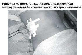 This is a manipulation that is carried out in a medical institution by a surgeon. For drainage of the liver, a skin puncture is performed at the site of the projection of the abscess. According to the doctor of functional diagnostics, puncture of the liver under ultrasound control gives the greatest control over the process. Next, the surgeon enters the abscess with a long needle and pumps out the pus with a syringe. Then, without removing the needle, an antiseptic solution (an agent that destroys bacteria) is drawn into the syringe and the abscess cavity is washed. After that, the patient is prescribed a course of antibiotic treatment. a wide range actions - cephalosporins, amcoxiclav, etc.
Useful sites about bile drainage
It is important that you discuss the results with the doctor who referred you in person or over the phone so they can explain what the results mean to you. Quality improvement recommendations for percutaneous transhepatic cholangiography and biliary drainage. Percutaneous transhepatic biliary drainage is a procedure indicated in patients with non-operative lesions when endoscopic application of a prosthesis is not possible due to anatomical reasons, complications, or the severe general condition of the patient. Most often, this is a palliative procedure aimed at improving the quality of life, although it does not change the prognosis of underlying diseases.
The consequence of incomplete drainage of the liver may be a recurrence of the disease and re-accumulation of pus. In rare cases, damage to a large vessel is possible during the insertion of a needle into a liver abscess - this is fraught with profuse bleeding. In general, drainage of the liver with a purulent abscess is definitely necessary, since not removed pus can lead to intoxication of the body and death of the patient.
Causes and symptoms of obstructive jaundice
This study presents our own experience of biliary drainage using the percutaneous transhepatic method and simultaneously evaluates the effectiveness and safety of the method. A total of 186 percutaneous drainage procedures were applied. Average age patients was 6 years. Bilirubin, alkaline phosphatase and gamma-glutamyltransferase were measured before and after the procedure. All data were analyzed statistically.
In the examined group, percutaneous drainage was successful in 7% of interventions. In 1% of the procedure, drainage application was ineffective. The most common complication during the procedure was hemobilia, and a long-term complication was sewage dislocation.
Another option for liver drainage is to clear the biliary tract of stagnant bile.
This is carried out with biliary dyskinesia caused by such reasons:
Viral hepatitis, especially chronic hepatitis;
Toxic damage to the liver (toxic hepatitis) caused by exposure to long-term medication, alcohol, fatty foods;
This technique is indicated in patients with neoplastic biliary obstruction with low expected survival and is thus a palliative procedure. A 40-year-old Hispanic woman presents to the emergency room with an acute right upper quadrant flank that started this morning and lasted over 2 hours. She woke up feeling both feverish and nauseous. She noticed that it was a continuous sharp, spasmodic pain, and she was more bothered when she urinated and saw that her urine was darker than usual.
Violations in the work of the autonomic nervous system, which leads to a violation of the excretion of bile from the biliary tract.
Technique
In a hospital, probing is carried out - a probe (thin tube) is inserted into the duodenum through the mouth, through which bile is excreted. The procedure is performed on an empty stomach, before probing the patient is given an antispasmodic drug. Under its action, the sphincter of the gallbladder relaxes and the output of bile improves. In addition to cutaneous, there are transabdominal methods. You can learn more about them in the article on liver puncture in more detail.
The patient also reported vomiting shortly after waking up. The patient denies pain on exertion or shortness of breath. She denies any recent respiratory infections or any extended travel. The patient is otherwise healthy with no significant past history illness. Past surgical history includes an uncomplicated elective C-section two years ago at the time of the birth of her second child. The family history is unremarkable, but the patient states that her mother had chronic cholelithiasis when she was in her fifties.
She is awake, awake and has no acute problems. The rest of the physical exam is unremarkable, except for pain on palpation of the right upper quadrant during the abdominal exam. Image of the correct upper quadrant to confirm the suspected diagnosis. Laboratory assessment.
Liver drainage at home is called blind probing or tubage. To use it, you need to drink 20 g of sorbitol dissolved in 1 glass of water at home in the morning on an empty stomach. Then you need to lie on your right side, placing a heating pad under the right hypochondrium (the area of the projection of the liver). Sorbitol has choleretic properties, and a heating pad in the liver area leads to relaxation of the sphincter of the gallbladder. As a result, stagnant bile is forced out of the bile ducts and the liver is drained. The course of such procedures is 3-5 times a day.
What is the most likely diagnosis? Bile duct disorder. Pathophysiology Blockade in any pathway that carries bile from the liver to the gallbladder or small intestine, which can lead to accumulation of both conjugated and unconjugated bilirubin and alkaline phosphatase. Bile: From cholesterol, bile salts and waste products. to the liver and cause obstructive biliary jaundice. Elevated levels of serum estrogen contribute to supersaturation of cholesterol in the bile. Elevated serum progesterone levels can lead to gallbladder congestion, promoting bile duct formation. Ursodeoxycholic acid prophylaxis has been shown to reduce stone formation during periods of rapid weight loss and is recommended for use in obese patients who are expected to lose large amounts of weight. Clofibrate: cholesterol-lowering agent; main side effect was production gallstones Ceftriaxone: secreted in bile and can be precipitated by calcium to form stones. Gastrointestinal tract: choledocholithiasis, acute or chronic pancreatitis Infection: cholangitis, parasitic infections Oncology: internal or external tumor dissection, lymphoma, pancreatic cancer, biliary cancer Malignant obstruction is usually associated with pancreatic carcinoma, cholangiocarcinoma, or metastatic disease. Symptoms include: Right upper quadrant abdominal pain Dark urine Fever Itching Jaundice Nausea and vomiting Pale-colored stools. What is one of the most common causes of malignant obstruction? bile ducts? Rapid weight loss Rapid use of female clofibrate Relatives American ancestry Increased physical activity. Biliary atresia Omphalocele Hirschsprung disease Primary sclerosing cholangitis Cholelithiasis. In the practice of interventional radiology. 1st ed.
- Life-threatening infection or sepsis.
- Diseases of the gallbladder and bile ducts.
- biliary intervention.
Consequences
In general, the consequences of such drainage are positive. A person after a course of blind probing feels much better, digestion and the functioning of the immune system improve.
However, if there are stones in the gallbladder, then hepatic colic can be provoked by drainage of the liver. Therefore, before undertaking such procedures, it is necessary to consult a doctor.
Ascites in cirrhosis of the liver
Against the background of morphological changes in the liver and hemodynamic and metabolic factors developing against their background, ascites develops. Ascites is an accumulation of free fluid in the abdominal cavity.
Liver recovery after antibiotics
The leading place in the development of acute inflammatory liver diseases (hepatitis) belongs to a viral infection, alcohol consumption, exposure to various industrial and natural poisons, as well as certain medications, including antibiotics.
The first signs of cirrhosis of the liver
Cirrhosis of the liver is a chronic progressive liver disease. This pathology is characterized by a decrease in the number of functioning hepatocytes (liver cells), a restructuring of the basic substance and the vascular system of the liver, and the development of liver failure and portal hypertension.
Hemangioma of the liver - causes and treatment
Hemangioma of the liver is called a benign tumor, the nature of which is heterogeneous. As a rule, the term hemangioma combines several varieties of vascular neoplasms of a blastomatous and disembryoplastic nature.
Drainage after surgery: the need and consequences
Surgery is the most conservative of the medical specialties. Methods, approaches to surgical treatment, the very rules for conducting operations have been verified over the years, and it takes a long time for any changes to become new rules. This is also the case with postoperative drainage.
Positional drainage during pregnancy
During pregnancy, at various times, most women experience health problems. But this rarely leads to a complete contraindication of gymnastics and is usually temporary. Properly selected exercises will help to carry out a better blood supply. internal organs, mainly the kidneys, will relieve lower back pain. The knee-elbow posture, for example, can be attributed to positional drainage, as it enhances the production and excretion of urine with toxins. In addition, by relieving pressure from the pelvic vessels, the outflow through the veins improves. lower extremities, which contributes to the treatment of edema or pastosity of the legs.