The organs of movement are a single system where each part and organ is formed and functions in constant interaction with each other. The elements included in the movement organ system are divided into two main categories: passive (bones, ligaments and joints) and active elements of the movement organs (muscles).
The size and shape of the human body is largely determined by the structural basis - the skeleton. The skeleton provides support and protection for the entire body and individual organs. The skeleton contains a system of movably articulated levers, driven by muscles, due to which various movements of the body and its parts are made in space. Individual parts of the skeleton serve not only as a container for vital organs, but also provide their protection. For example, the skull, chest and pelvis serve as protection for the brain, lungs, heart, intestines, etc.
Until recently, the prevailing opinion was that the role of the skeleton in the human body is limited to the function of supporting the body and participation in movement (this was the reason for the appearance of the term “musculoskeletal system”). Thanks to modern research, the understanding of the functions of the skeleton has expanded significantly. For example, the skeleton is actively involved in metabolism, namely in maintaining the mineral composition of the blood at a certain level. Substances that make up the skeleton, such as calcium, phosphorus, citric acid and others, easily enter into metabolic reactions if necessary. The function of muscles is also not limited to the inclusion of bones in movement and performance of work; many muscles, surrounding the body cavities, protect internal organs.
General information about the skeleton. Bone Shape
The human skeleton is similar in structure to the skeleton of higher animals, but has whole line features associated with upright walking, walking on two limbs, high development hands and brain.
The human skeleton is a system consisting of 206 bones, of which 85 are paired and 36 are unpaired. Bones are organs of the body. The weight of the skeleton in a man is approximately 18% of body weight, in a woman - 16%, in a newborn - 14%. The skeleton consists of bones of various sizes and shapes.
According to their shape, bones are divided into:
A) long (located in the skeleton of the limbs);
b) short (located in the wrist and tarsus, i.e., where greater strength and mobility of the skeleton are simultaneously required);
V) wide or flat (they form the walls of cavities in which internal organs are located - the pelvic bone, the bones of the skull);
G) mixed (have different shapes).
Bone connections
Bones articulate different ways. According to the degree of mobility, joints are distinguished: a) fixed; b) sedentary; c) movable bone joints, or joints.
A fixed joint is formed as a result of bone fusion, and movement may be extremely limited or completely absent. For example, the immobility of the bones of the brain skull is ensured by the fact that numerous protrusions of one bone fit into the corresponding depression of another. This connection of bones is called a suture.
The presence of elastic cartilage pads between the bones provides slight mobility. For example, such gaskets exist between individual vertebrae. During muscle contraction, the pads compress and the vertebrae move closer together. During active movements (walking, running, jumping), cartilage acts as a shock absorber, thereby softening sharp shocks and protecting the body from shaking.
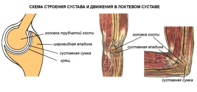
Movable bone connections are more common, which is provided by joints. The ends of the bones forming the joint are covered with hyaline cartilage with a thickness of 0.2 to 0.6 mm. This cartilage is very elastic, has a smooth shiny surface, therefore the friction between the bones is significantly reduced, which greatly facilitates their movement.
A very dense connective tissue forms the joint capsule (capsule), which surrounds the area of bone articulation. The strong outer (fibrous) layer of the capsule firmly connects the articulating bones to each other. The inside of the capsule is lined with synovial membrane. The joint cavity contains synovial fluid, which acts as a lubricant and also helps reduce friction.
The outside of the joint is strengthened by ligaments. A number of joints are strengthened by ligaments and internally. In addition, inside the joints there are special devices that increase the articulated surfaces: lips, discs, menisci made of connective tissue and cartilage.
The joint cavity is hermetically sealed. The pressure between the articular surfaces is always negative (less than atmospheric), and therefore external atmospheric pressure prevents their divergence.
Types of joints
According to the shape of the articular surface and the axes of rotation, joints are distinguished:
A) with three;
b) with two;
V) with one axis of rotation.
The first group consists of spherical joints - the most mobile (for example, the joint between the scapula and humerus). The joint between the innominate bone and the femur, called the nut joint, is a type of ball and socket joint.
The second group consists of ellipsoidal (for example, the joint between the skull and the first cervical vertebra) and saddle joints (for example, the joint between the metacarpal bone of the first finger and the corresponding bone of the wrist).
The third group includes trochlear joints (joints between the phalanges of the fingers), cylindrical joints (between the ulna and radius bones) and helical joints (forming the elbow joint).
Any unfixed body has six degrees of freedom, because it produces three translational and three rotational movements along the coordinate axes. A fixed body can only produce rotations. Since all the links of the body are fixed, joints with three axes of rotation are the most mobile and have three degrees of freedom. Joints with two axes of rotation are less mobile and therefore have two degrees of freedom. Joints with one axis of rotation have one degree of freedom, which means the least mobility.
Bone structure
Each bone is a complex organ consisting of bone tissue, periosteum, bone marrow, blood and lymphatic vessels and nerves. With the exception of the connecting surfaces, the entire bone is covered with periosteum - a thin connective tissue membrane rich in nerves and vessels that penetrate from it into the bone through special openings. Ligaments and muscles are attached to the periosteum. The cells that make up the inner layer of the periosteum grow and multiply, which ensures the growth of the bone in thickness, and in the event of a fracture, the formation of a callus.
By sawing a tubular bone along the long axis, you can see that there is a dense (or compact) bone substance on the surface, and a spongy substance underneath (in depth). In short bones, such as the vertebrae, spongy material predominates. Depending on the load the bone experiences, the compact substance forms a layer of varying thickness. The cancellous substance is formed by very thin bone bars oriented parallel to the main stress lines. This allows the bone to withstand significant loads.
The dense layer of bone has a lamellar structure and is similar to a system of cylinders inserted into each other, which also gives the bone strength and lightness. Between the plates of bone substance lie bone tissue cells. Bone plates make up the intercellular substance of bone tissue.
Tubular bone consists of a body (diaphysis) and two ends (epiphyses). On the epiphyses there are articular surfaces that are covered with cartilage, which is involved in the formation of the joint. On the surface of the bones there are tubercles, tubercles, grooves, ridges, notches to which muscle tendons are attached, as well as openings through which blood vessels and nerves pass.
Chemical composition of bone
Dried and defatted bone has the following composition: organic matter – 30%; minerals – 60%; water – 10%.
The organic substances of bone include fibrous protein (collagen), carbohydrates and many enzymes.
Bone minerals are represented by salts of calcium, phosphorus, magnesium and many trace elements (such as aluminum, fluorine, manganese, lead, strontium, uranium, cobalt, iron, molybdenum, etc.). The adult human skeleton contains about 1200 g of calcium, 530 g of phosphorus, 11 g of magnesium, i.e. 99% of all calcium present in the human body is contained in the bones.
In children, organic substances predominate in the bone tissue, so their skeleton is more flexible, elastic, and easily deformed under prolonged and heavy loads or incorrect body positions. The amount of minerals in bones increases with age, causing bones to become more fragile and more likely to break.
Organic and mineral substances make bone strong, hard and elastic. The strength of the bone is also ensured by its structure, the location of the bone crossbars of the spongy substance according to the direction of the pressure and tensile forces.
Bone is 30 times harder than brick, granite - 2.5 times. Bone is stronger than oak. It is nine times stronger than lead and almost as strong as cast iron. In a vertical position, the human femur can withstand load pressure of up to 1500 kg, and tibia– up to 1800 kg.
Development skeletal system in childhood and adolescence
During prenatal development in children, the skeleton consists of cartilage tissue. Ossification points appear after 7–8 weeks. The newborn has ossified diaphysis of the tubular bones. After birth, the ossification process continues. The timing of the appearance of ossification points and the end of ossification varies for different bones. Moreover, for each bone they are relatively constant; they can be used to judge the normal development of the skeleton in children and their age.
The skeleton of a child differs from the skeleton of an adult in its size, proportions, structure and chemical composition. Skeletal development in children determines the development of the body (for example, muscles develop more slowly than the skeleton grows).
There are two ways of bone development.
1. Primary ossification, when bones develop directly from embryonic connective tissue - mesenchyme (bones of the calvarium, facial part, partly the clavicle, etc.). First, a skeletogenic mesenchymal syncytium is formed. It contains cells - osteoblasts, which turn into bone cells - osteocytes, and fibrils, impregnated with calcium salts and turning into bone plates. Thus, bone develops from connective tissue.
2. Secondary ossification, when bones are initially laid down in the form of dense mesenchymal formations, having the approximate outlines of future bones, then turn into cartilage tissue and are replaced by bone tissue (bones of the base of the skull, torso and limbs).
With secondary ossification, the development of bone tissue occurs by replacement both externally and internally. Externally, the formation of bone substance occurs by osteoblasts of the periosteum. Internally, ossification begins with the formation of ossification nuclei, and gradually the cartilage is resorbed and replaced by bone. As the bone grows, it is absorbed from the inside by special cells - osteoclasts. Bone growth occurs from the outside. Bone growth in length occurs due to the formation of bone substance in the cartilage located between the epiphysis and diaphysis. These cartilages gradually move towards the epiphysis.
Many bones in the human body are not formed entirely, but in separate parts, which then merge into a single bone. For example, the pelvic bone first consists of three parts that fuse together by the age of 14–16 years. Tubular bones are also formed in three main parts (the ossification nuclei in the places where bony protrusions are formed are not taken into account). For example, the fetal tibia initially consists of solid hyaline cartilage. Ossification begins in the middle part at approximately the eighth week of intrauterine life. Replacement of the diaphysis with bone occurs gradually and occurs first from the outside and then from the inside. In this case, the epiphyses remain cartilaginous. The ossification nucleus in the upper epiphysis appears after birth, and in the lower epiphysis - in the second year of life. In the middle part of the epiphyses, the bone first grows from the inside, then from the outside, as a result of which two layers of epiphyseal cartilage remain separating the diaphysis from the epiphyses.
In the upper epiphysis of the femur, the formation of bone beams occurs at the age of 4–5 years. After 7–8 years they lengthen and become uniform and compact. The thickness of the epiphyseal cartilage reaches 2–2.5 mm by the age of 17–18 years. By the age of 24, the growth of the upper end of the bone ends and the upper epiphysis fuses with the diaphysis. The lower epiphysis grows to the diaphysis even earlier - by 22 years. With the end of ossification of the tubular bones, their growth in length stops.
Ossification process
General ossification of the tubular bones is completed by the end of puberty: in women - by 17-21 years, in men - by 19-24 years. Because men reach puberty later than women, they are taller on average.
From five months to one and a half years, i.e., when the child stands on his feet, the main development of the lamellar bone occurs. By 2.5–3 years, the remains of coarse fibrous tissue are no longer present, although during the second year of life most of the bone tissue has a lamellar structure.
Reduced function of the endocrine glands (anterior part of the adenohypophysis, thyroid, parathyroid, thymus, sex) and a lack of vitamins (especially vitamin D) can cause delayed ossification. Acceleration of ossification occurs with premature puberty, increased function of the anterior part of the adenohypophysis, thyroid gland and adrenal cortex. Delayed and accelerated ossification most often manifests itself before the age of 17–18 years, and the difference between “bone” and passport ages can reach 5–10 years. Sometimes ossification occurs faster or slower on one side of the body than on the other.
As we age, the chemical composition of our bones changes. Children's bones contain more organic matter and less inorganic matter. As it grows, the amount of calcium salts, phosphorus, magnesium and other elements increases significantly, and the ratio between them changes. Thus, in young children, calcium is retained most in the bones, but as they grow older, there is a shift towards greater phosphorus retention. Inorganic substances in the bones of a newborn make up one-second of the weight of the bone, in an adult - four-fifths.
Change of structure and chemical composition changes in bones also entail a change in their physical properties. Children's bones are more elastic and less brittle than adults'. Cartilage in children is also more flexible.
Age-related differences in the structure and composition of bones are especially clearly manifested in the number, location and structure of the Haversian canals. With age, their number decreases, and their location and structure change. How older child, the more dense substance there is in his bones; small children have more spongy substance. By the age of 7, the structure of the tubular bones is similar to that of an adult, however, between 10–12 years, the spongy substance of the bones changes even more intensively, its structure stabilizes by 18–20 years.
How younger child, the more the periosteum is fused to the bone. The final differentiation between bone and periosteum occurs by the age of 7 years. By the age of 12, the dense substance of the bone has an almost uniform structure; by the age of 15, single areas of resorption of the dense substance completely disappear, and by the age of 17, large osteocytes predominate in it.
From 7 to 10 years, the growth of the bone marrow cavity in the tubular bones slows down sharply, and it is finally formed from 11–12 to 18 years. The enlargement of the medullary canal occurs in parallel with the uniform growth of the dense substance.
Between the plates of the spongy substance and in the medullary canal there is bone marrow. Due to big amount blood vessels in the tissues of newborns there is only red bone marrow - hematopoiesis occurs in it. At six months, the gradual process of replacing red bone marrow in the diaphysis of the tubular bones with yellow bone marrow, consisting mostly of fat cells, begins. Replacement of the red brain ends by the age of 12–15 years. In adults, red bone marrow is stored in the epiphyses of long bones, in the sternum, ribs and spine and amounts to approximately 1500 cubic meters. cm.
The healing of fractures and the formation of callus in children occurs after 21–25 days; in infants this process occurs even faster. Dislocations in children under 10 years of age are rare due to the high extensibility of the ligamentous apparatus.
Approximately every twentieth person has osteoarthritis, every tenth person experiences it regularly, and more than 70% of the population experiences it from time to time or sporadically. Problems with the musculoskeletal system are so common mainly due to an irresponsible attitude towards this aspect, while preventive measures require almost no special effort.
What is this
The human musculoskeletal system is a systemically interconnected set of bones (forming the skeleton) and their joints, allowing a person to control (through impulses transmitted through the nervous system by the brain) the body, its statics and dynamics. The importance of the human musculoskeletal system is difficult to overestimate. A person whose ODS does not fulfill its functions is, at best, a disabled person or a paralytic lying flat.
Did you know? One of the founders of anatomy in its modern, scientific form was Leonardo da Vinci. He, along with other Renaissance scientists and researchers, performed autopsies to understand the structure of the human body.
In a healthy person, the functions of the musculoskeletal system are divided into mechanical and biological.
Basic Mechanical Functions
Mechanical functions are associated with the preservation of the structure and movement of the body in space.
Support
It consists of forming the basis for the rest of the body - muscles, tissues and organs are attached to the skeleton. Due to the skeleton and the muscles attached to it, a person can stand upright, his organs maintain a relatively static position relative to the axis of symmetry and each other.
Protective
Bones protect the most important internal organs from mechanical damage: the head is protected by the skull, the back is protected by the spine, the internal organs of the chest (, lungs and others) are hidden behind the ribs, the genitals are covered by the pelvic bones.  It is this kind of protection that provides us with resistance to external influences, and well-trained muscles can enhance this effect.
Did you know? At the moment of our birth, we have the most bones - 300. Subsequently, some fuse (and all become stronger) and their total number decreases to 206.
Motor
The most prominent function of the human musculoskeletal system. The building muscles are attached to the skeleton. Due to their contractions, various movements are performed: flexion/extension of limbs, walking and much more.
Actually, this is one of the main differences between representatives of the biological kingdom “Animals” - conscious and controlled movements in space.
Spring
Softening (cushioning) of movements due to the structure and position of bones and cartilage.  It is provided both by the shape of the bones (for example, the bend of the foot, strong shin bones - an evolutionary mechanism that is most suitable for walking upright and supporting body weight with an emphasis on only one pair of limbs), and by auxiliary tissues - cartilage and joint capsules ensure a decrease in friction of the bones in their places joints.
Biological functions of the system
The musculoskeletal system also has other functions that are important for life.
Hematopoietic
The process of blood formation occurs in the so-called red bone marrow, but due to its location (in the tubular bones), this function is also classified as ODA.
In the red bone marrow, hematopoiesis (blood formation) occurs - the creation of new blood cells, and partially immunopoiesis - the maturation of cells that take part in the functioning of the immune system.
Storage
A large amount of substances necessary for the body, such as, and, accumulate and store in the bones. From there they flow to other organs, where they are included in the metabolic process. 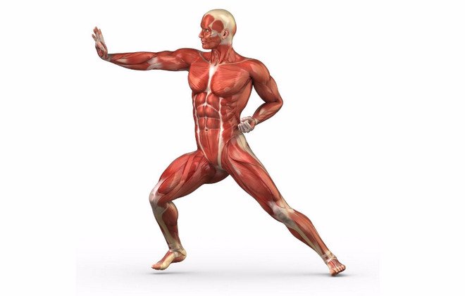 These substances ensure the strength of bones and their resistance to external influences, as well as the speed of healing after fractures.
Important! Problems with calcium are often caused not by insufficient intake, but by rapid “leaching out”. This is facilitated by such popular products as sweet carbonated drinks and oxalic acid. It is better to exclude all this from the diet.
Main problems and injuries of the musculoskeletal system
Although the formation of the musculoskeletal system occurs in, its development is a process that continues throughout.
The causes of problems with ODA, as well as their consequences, can be different:- Incorrect load (insufficient or excessive).
- Inflammatory processes affecting bone tissue, muscle or cartilage. Depending on the etiology and location, the diagnosis varies.
- Disorders associated with metabolism, deficiency or excess of any elements.
- Mechanical injuries (bruises, fractures) and the consequences of improper treatment.
Diseases of the musculoskeletal system
Diseases affecting our musculoskeletal system are depressing in their diversity:
- Arthritis affects the joints and can develop into arthrosis.
- Infections can settle in the periarticular bursa (bursitis), muscles (myotitis), bone marrow (osteomyelitis), and large joints (periarthritis).
- The spine may bend, the ankle may lose tone.
Important! For any pain, consult a doctor! In the early stages of the disease, ODA is treated with simple and gentle methods: physical or manual therapy, medical therapy. If the disease is in a severe stage, treatment and rehabilitation will be long and difficult.
Sports injuries
Of course, with the right amount of “luck”, you can fall out of the blue and at the same time break something unexpected.
However, according to statistics, the most common injuries during sports are: muscle strains, various injuries to the lower leg, fractures (mainly the legs) and ruptures (of ligaments, cartilage or tendons). 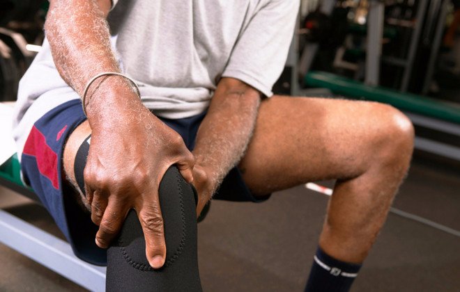
Staying healthy: how to prevent troubles
To keep the body in good shape and the musculoskeletal system in working and healthy condition, it is important to know what measures to take to maintain normal functions of the musculoskeletal system.
Nothing fancy is required:
- Healthy lifestyle.
- A balanced diet rich in calcium and other minerals and trace elements.
- Regular physical activity appropriate for age and health.
- Walks in the sun (vitamin D) and fresh air.
- Maintaining optimal body weight (obesity, like dystrophy, are the enemies of the musculoskeletal system).
- Convenient workplace.
- Regular medical checkups.
As you can see, if you support the body as a whole, its systems will also be fine. You don't have to play sports professionally to do this. 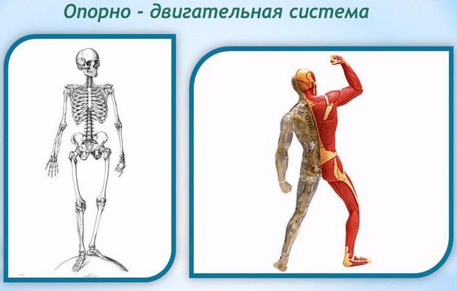 It will be enough not to neglect physical activity (in any form convenient for you, be it yoga, swimming or regular walks in the park), follow a daily routine and maintain a healthy diet. It is not so difficult. Do not be ill!
Consists of a skeleton and muscles, it performs the following functions:
Protective (limits the cavities in which the internal organs are located);
Support function;
Provides active human movements;
Performs a hematopoietic function;
Participates in metabolism.
The passive part of the musculoskeletal system is the skeleton, consisting of bones, cartilage, joints and ligaments. There are more than 200 bones in the human skeleton.
Each bone is an organ consisting of bone tissue.
Bone= cells with processes + intercellular substance + nerves + vessels + connective tissue membrane
Bones:
(bone properties): organic substances (flexibility and elasticity), inorganic substances (hardness).
Direction of growth (source of new cells): in length (cartilage), in thickness (periosteum).
Bone connection: movable, semi-movable, immobile
Joint– articulating bone with glenoid cavity + articulating bone with head + strong ligaments + articular capsule + articular fluid
Human skeleton consists of 200 bones.
Main departments:
Muscles– an active part of the musculoskeletal system, providing all the variety of movements performed in the human body. Thanks to the muscles, the body maintains balance, moves in space, breathing movements are carried out with the chest and diaphragm, swallowing, the voice is formed, eye movements are carried out, and internal organs work, including the heart. Humans have two types of muscles: smooth and striated.
Smooth muscles are found in internal organs: the walls of blood vessels, the bladder, ureters, and intestines. Their reduction occurs arbitrarily.
Striated muscles provide muscle attachment to the tendons and bones of the skeleton. Skeletal muscles move bones relative to each other in compositions; in addition, they participate in the formation of the walls of the abdominal and thoracic cavities and pelvis. They form part of the wall of the upper part of the esophagus and larynx. Carry out the movement of the apple, breathing and swallowing movements. All skeletal muscles can be divided into two groups - flexors and extensors.
Facial muscles are facial muscles that are not connected to joints.
The heart muscle is a special striated muscle, where the fibers are connected, and contracts rapidly.
In humans, each muscle contains all types of muscle fibers; their ratio varies depending on the purpose of each muscle. Each muscle is approached by blood vessels that penetrate the outer shell and break up into a network of capillaries in the muscle. Blood supplies muscle fibers with oxygen and nutrients. In addition, each muscle has a nerve that transmits signals.
Abstract on biology on the topic:
"Musculoskeletal system"
Pupil 9 "G" class
high school № 117
SWAD Moscow
Yuditsky Alexander.
Moscow 2004
Plan:
I. Introduction.
II. Skeleton.
1. Spine.
2. Chest.
3.Limbs.
4. Leg and arm.
III. Two types of muscle tissue.
1. Smooth muscles.
2. Skeletal muscles.
3. Nerve connections in muscles.
4.Muscles produce heat.
5.Strength and speed of muscle contraction.
IV. Fatigue and rest.
1. Causes of fatigue.
V. Statics and dynamics of the human body.
1. Equilibrium conditions.
VI. Everyone needs sports.
1.Muscle training.
2.Work and sports.
3. Anyone can become an athlete.
VII.
VIII. Conclusion.
XI.
Musculoskeletal system
The musculoskeletal system consists of skeletal bones with joints, ligaments and muscles with tendons, which, along with movements, provide the supporting function of the body. Bones and joints participate in movement passively, subject to the action of muscles, but play a leading role in the implementation of the support function. The specific shape and structure of the bones gives them greater strength, the reserve of which for compression, expansion, and flexion significantly exceeds the loads possible during the daily work of the musculoskeletal system. For example, the human tibia, when compressed, can withstand a load of more than a ton, and in terms of tensile strength it is almost as good as cast iron. Ligaments and cartilage also have a large margin of strength.
The skeleton consists of bones connected to each other. It provides our body with support and shape, and also protects our internal organs. An adult human skeleton consists of approximately 200 bones. Each bone has a certain shape, size and occupies a certain position in the skeleton. Some of the bones are connected to each other by movable joints. They are driven by muscles attached to them.
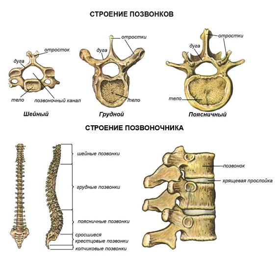
Spine. The original structure that forms the main support of the skeleton is the spine. If it consisted of a solid bone rod, then our movements would be constrained, lacking flexibility and would cause just as much pain. discomfort like riding in a cart without springs on a cobblestone street.
The elasticity of hundreds of ligaments, cartilage layers and bends makes the spine a strong and flexible support. Thanks to this structure of the spine, a person can bend, jump, somersault, and run. Very strong intervertebral ligaments allow the most complex movements and at the same time create reliable protection for the spinal cord. It is not subjected to any mechanical stretching or pressure during the most incredible bends of the spine.
The bends of the spinal column correspond to the influence of the load on the skeletal axis. Therefore, the lower, more massive part becomes a support when moving; The upper one, with free movement, helps maintain balance. The spinal column could be called the vertebral spring.
The wavy curves of the spine ensure its elasticity. They appear with the development of the child’s motor abilities, when he begins to hold his head up, stand, and walk.
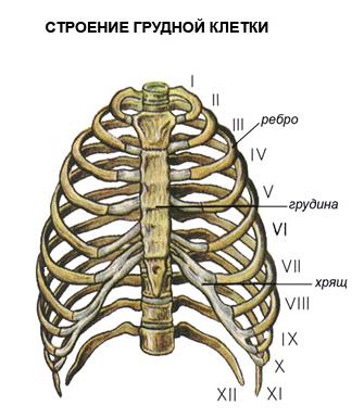
Rib cage. The rib cage is formed by the thoracic vertebrae, twelve pairs of ribs and a flat breastbone, or breastbone. The ribs are flat, curved bones. Their posterior ends are movably connected to the thoracic vertebrae, and the anterior ends of the ten upper ribs are connected to the sternum with the help of flexible cartilage. This ensures the mobility of the chest during breathing. The two lower pairs of ribs are shorter than the others and end freely. The rib cage protects the heart and lungs, as well as the liver and stomach.
It is interesting to note that ossification of the chest occurs later than other bones. By the age of twenty, the ossification of the ribs ends, and only by the age of thirty does the complete fusion of the parts of the sternum, consisting of the manubrium, the body of the sternum and the xiphoid process, occur.
The shape of the chest changes with age. In a newborn, it usually has the shape of a cone with the base facing down. Then, in the first three years, the circumference of the chest increases faster than the length of the body. Gradually, the chest from a cone-shaped one acquires a round shape characteristic of a person. Its diameter is greater than its length.
The development of the chest depends on a person’s lifestyle. Compare an athlete, swimmer, athlete with a person who does not play sports. It is easy to understand that the development of the chest and its mobility depend on the development of muscles. Therefore, teenagers twelve to fifteen years old who play sports have a chest circumference that is seven to eight centimeters larger than their peers who do not play sports.
Incorrect seating of students at their desks and compression of the chest can lead to its deformation, which impairs the development of the heart, large vessels and lungs.
Limbs. Due to the fact that the limbs are attached to a reliable support, they have mobility in all directions and are able to withstand heavy physical loads.
Light bones - collarbones and shoulder blades, lying on the upper part of the chest, cover it like a belt. This is the support of the hands. The projections and ridges on the collarbone and scapula are where the muscles attach. The greater the strength of these muscles, the more developed the bone processes and irregularities. In an athlete or a loader, the longitudinal ridge of the scapula is more developed than in a watchmaker or accountant. The collarbone is a bridge between the bones of the torso and arms. The shoulder blade and collarbone create a reliable spring support for the arm.
The position of the arms can be judged by the position of the shoulder blades and collarbones. Anatomists have helped restore the broken arms of an ancient Greek statue of the Venus de Milo, determining their position based on the silhouettes of the shoulder blades and collarbones.
The pelvic bones are thick, wide and almost completely fused. In humans, the pelvis lives up to its name - it, like a bowl, supports the internal organs from below. This is one of the typical features of the human skeleton. The massiveness of the pelvis is proportional to the massiveness of the leg bones, which bear the main load when a person moves, so the human pelvic skeleton can withstand a large load.
Leg and arm. With a vertical posture, a person’s hands do not bear a constant load as a support, they acquire ease and variety of action, and freedom of movement. The hand can perform hundreds of thousands of different motor operations. The legs bear the entire weight of the body. They are massive and have extremely strong bones and ligaments.
The head of the shoulder has no restrictions in wide circular movements of the arms, for example when throwing a javelin. The head of the femur protrudes deeply into the socket of the pelvis, which limits movement. The ligaments of this joint are the strongest and hold the weight of the body on the hips.
Through exercise and training, greater freedom of movement of the legs is achieved, despite their massiveness. A convincing example of this can be ballet, gymnastics, and martial arts.
The tubular bones of the arms and legs have a huge margin of strength. It is interesting that the arrangement of the openwork crossbars of the Eiffel Tower corresponds to the structure of the spongy substance of the heads of the tubular bones, as if J. Eiffel designed the bones. The engineer used the same laws of construction that determine the structure of bone, giving it lightness and strength. This is the reason for the similarity between the metal structure and the living bone structure.
The elbow joint provides complex and varied movements of the arm in a person’s working life. Only he has the ability to rotate the forearm around its axis, with a characteristic movement of unwinding or twisting.
Knee-joint guides the lower leg when walking, running, jumping. The knee ligaments in humans determine the strength of the support when straightening the limb.
The hand begins with a group of carpal bones. These bones do not experience strong pressure and perform a similar function, so they are small, uniform, and difficult to distinguish. It is interesting to mention that the great anatomist Andrei Vesalius could, blindfolded, identify each carpal bone and say whether it belonged to the left or right hand.
The bones of the metacarpus are moderately mobile, they are arranged in the form of a fan and serve as a support for the fingers. Phalanges of fingers - 14. All fingers have three bones, except the thumb - it has two bones. A person's thumb is very mobile. It can become at right angles to everyone else. Its metacarpal bone is capable of opposing the rest of the bones of the hand.
The development of the thumb is associated with labor movements of the hand. The Indians call the thumb “mother”, the Javanese call it “elder brother”. In ancient times, captives had their thumbs cut off to degrade their human dignity and render them unfit to fight.
The brush makes the most subtle movements. In any working position of the hand, the hand retains complete freedom of movement.
The foot became more massive due to walking. The tarsal bones are very large and strong compared to the carpal bones. The largest of them are the talus and calcaneus. They can withstand significant body weight. In newborns, the movements of the foot and its big toe are similar to their movement in monkeys. Strengthening the supporting role of the foot when walking led to the formation of its arch. When walking or standing, you can easily feel how the entire space between these points “hangs in the air.”
The vault, as is known in mechanics, can withstand greater pressure than the platform. The arch of the foot ensures elasticity of gait and eliminates pressure on nerves and blood vessels. His education in the history of human origins is associated with upright walking and is distinctive feature of a person acquired in the process of his historical development.
Two types of muscle tissue.
Smooth muscles. When we talked about muscles, we usually thought of skeletal muscles. But, besides them, in our body in the connective tissue there are smooth muscles in the form of single cells, in some places they are collected in bundles.
There are many smooth muscles in the skin, they are located at the base of the hair follicle. By contracting, these muscles lift the hair and squeeze out fat from the sebaceous gland.
In the eye around the pupil there are smooth circular and radial muscles. They work all the time, unbeknownst to us: in bright light the circular muscles constrict the pupil, and in the dark the radial muscles contract and the pupil dilates.
In the walls of all tubular organs - respiratory tract, blood vessels, digestive tract, urethra, etc. - there is a layer of smooth muscle. Under the influence of nerve impulses it contracts. For example, its contraction in the windpipe delays the flow of air containing harmful impurities - dust, gases.
Due to the contraction and relaxation of smooth cells in the walls of blood vessels, their lumen either narrows or expands, which contributes to the distribution of blood in the body. The smooth muscles of the esophagus contract and push a bolus of food or a sip of water into the stomach.
Complex plexuses of smooth muscle cells are formed in organs with a wide cavity - in the stomach, bladder, uterus. The contraction of these cells causes compression and narrowing of the organ lumen. The force of each contraction of the cells is negligible because they are very small. However, the addition of the forces of entire bundles can create a contraction of enormous force. Powerful contractions create a sensation of intense pain.
Skeletal muscles. Skeletal muscles carry out both static activity, fixing the body in a certain position, and dynamic activity, ensuring the movement of the body in space and its individual parts relative to each other. Both types of muscle activity closely interact, complementing each other: static activity provides a natural background for dynamic activity. As a rule, the position of the joint is changed with the help of several muscles of multidirectional, including opposite action. Complex joint movements are performed by coordinated, simultaneous or sequential contraction of non-directional muscles. Consistency (coordination) is especially necessary to perform motor acts in which many joints are involved (for example, skiing, swimming).
Skeletal muscles are not only the executive motor apparatus, but also unique sensory organs. Muscle fibers and tendons contain nerve endings - receptors that send impulses to cells at various levels of the central nervous system. As a result, a closed cycle is created: impulses from various formations of the central nervous system, traveling along the motor nerves, cause muscle contraction, and impulses sent by muscle receptors inform the central nervous system about each element of the system. The cyclic system of connections ensures precision of movements and their coordination. Although the movement of skeletal muscles is controlled by various sections of the central nervous system, the leading role in ensuring interaction and setting the goal of a motor reaction belongs to the cerebral cortex. In the cerebral cortex, the motor and sensory zones of the representations form a single system, with each muscle group corresponding to a certain section of these zones. This relationship allows you to perform movements, attributing them to environmental factors acting on the body. Schematically, the control of voluntary movements can be represented as follows. The tasks and purpose of a motor action are formed by thinking, which determines the direction of a person’s attention and efforts. Thinking and emotions accumulate and direct these efforts. The mechanisms of higher nervous activity form the interaction of psychophysiological mechanisms of movement control at various levels. Based on the interaction of the musculoskeletal system, the development and correction of motor activity is ensured. Analyzers play a major role in the implementation of motor reactions. The motor analyzer ensures the dynamics and interrelation of muscle contractions and participates in the spatial and temporal organization of the motor act. The balance analyzer, or vestibular analyzer, interacts with the motor analyzer when the body position in space changes. Vision and hearing, actively perceiving information from the environment, are involved in spatial orientation and correction of motor reactions.
The name “muscle” comes from the word “musculis”, which means “mouse”.
This is due to the fact that anatomists, observing the contraction of skeletal muscles, noticed that they seemed to be running under the skin, like mice.
The muscle consists of muscle plexuses. The length of muscle plexuses in humans reaches 12 cm. Each such plexus forms a separate muscle fiber.
Under the muscle fiber sheath there are numerous rod-shaped nuclei. Along the entire length of the cell stretches several hundred thin strands of cytoplasm - myofibrils, capable of contracting. In turn, myofibrils are formed by 2.5 thousand protein threads.
The myofibrils alternate between light and dark discs, and under a microscope the muscle fiber appears transversely striated. Let's compare the function of skeletal and smooth muscles. It turns out that striated muscles cannot lengthen as much as smooth muscles. But skeletal muscles contract faster than the muscles of internal organs. It is therefore not difficult to explain why a snail or earthworm, lacking striated muscles, moves slowly. The rapidity of movements of a bee, lizard, eagle, horse, and human is ensured by the speed of contraction of the striated muscles.
The thickness of muscle fibers varies from person to person. For those who play sports, muscle fibers develop well, their mass is large, which means the force of contraction is also large. Limitation of muscle work leads to a significant reduction in the thickness of fibers and muscle mass as a whole, which also leads to a decrease in the force of contraction.
There are a total of 656 skeletal muscles in the human body. Almost all muscles are paired. The position of the muscles, their shape, and the method of attachment to the bones have been studied in detail by anatomy. The location and structure of the muscles is especially important for the surgeon to know. That is why the surgeon is first and foremost an anatomist, and anatomy and surgery are sisters. World achievements in the development of these sciences belong to our domestic science, and first of all to N.I. Pirogov.
Nerve connections in muscles. It is wrong to think that a muscle can contract on its own. It would be difficult to imagine a single coordinated movement if the muscles were uncontrollable. Nerve impulses “set off” the muscle. One muscle receives an average of 20 impulses per second. Each step, for example, involves up to 300 muscles, and many impulses coordinate their work.
The number of nerve endings in different muscles is not the same. There are relatively few of them in the thigh muscles, and the oculomotor muscles, which perform subtle and precise movements all day long, are rich in motor nerve endings. The hemispheric cortex is unevenly connected to individual muscle groups. For example, large areas of the cortex are occupied by motor areas that control the muscles of the face, hand, lips, and feet, and relatively small areas by the muscles of the shoulder, thigh, and lower leg. The size of individual zones of the motor cortex is proportional not to the mass of muscle tissue, but to the subtlety and complexity of the movements of the corresponding organs.
Each muscle has a double nerve subordination. One nerve carries impulses from the brain and spinal cord. They cause muscle contraction. Others, moving away from the nodes that lie on the sides of the spinal cord, regulate their nutrition.
The nerve signals that control movement and nutrition of muscles are consistent with the nervous regulation of blood supply to the muscle. This results in a single triple nervous control.
Muscles produce heat. Striated muscles are “engines” in which chemical energy is converted directly into mechanical energy. The muscle is used for movement 33% chemical energy, which is released during the breakdown of animal starch - glycogen. 67% of the energy in the form of heat is transferred by the blood to other tissues and uniformly warms the body. That is why in the cold a person tries to move more, as if warming himself up using the energy generated by the muscles. Small involuntary muscle contractions cause trembling - the body increases heat production.
Strength and speed of muscle contraction. The strength of a muscle depends on the number of muscle fibers, its cross-sectional area, the size of the bone surface to which it is attached, the angle of attachment and the frequency of nerve impulses. All these factors have been identified by special research.
The strength of a person's muscles is determined by how much weight he can lift. Muscles outside the body develop a force several times greater than that which is manifested in human movements.
The working quality of a muscle is associated with its ability to suddenly change its elasticity. When contracted, muscle protein becomes very elastic. After muscle contraction, it again acquires its original state. Becoming elastic, the muscle holds the load, and muscle strength is manifested in this. Human muscles develop a force of up to 156.8 N for each square centimeter of cross-section.
One of the most strong muscles– calf. It can lift a load of 130 kg. Every healthy person is able to “stand on tiptoes” on one leg and even lift an additional load. This load falls mainly on the calf muscle.
Being under the influence of constant nerve impulses, the muscles of our body are always tense, or, as they say, are in a state of tone - a prolonged contraction. You can test muscle tone on yourself: close your eyes with force, and you will feel the trembling of the contracted muscles in the eye area.
It is known that any muscle can contract with different forces. For example, the same muscles are involved in lifting a small stone and a pound weight, but they expend different forces. The speed at which we can move our muscles varies and depends on the training of the body. A violinist makes 10 movements per second, and a pianist makes up to 40.
Fatigue and rest
Causes of fatigue. Fatigue is an indicator that the body cannot work at full capacity. Why does muscle fatigue occur? For science, this question was unresolved for a long time. Various theories were created.
Some scientists assumed that the muscle was depleted from a lack of nutrients; Others said that she was “suffocating”, lack of oxygen. It has been suggested that fatigue occurs due to poisoning, or clogging, of the muscle with toxic waste products. However, all these theories did not satisfactorily explain the causes of fatigue. As a result, the assumption arose that the cause of fatigue did not lie in the muscle. Nerve fatigue was hypothesized. However, the outstanding Russian physiologist, one of I.M. Sechenov’s students, Professor N.E. Vvdensky, used an example to prove that nerve conductors are practically not fatigued.
The path to solving the mystery of fatigue was opened by the Russian physiologist I.M. Sechenov. He developed the nervous theory of fatigue. He found that the right hand, after prolonged work, restored its functionality if movements were made with the left hand during the rest period. The nerve centers of the left hand seemed to charge the tired nerve centers of the right hand with energy. It turned out that fatigue is relieved more quickly when the rest of the working hand is combined with the work of the other hand than with complete rest. With these experiments, I.M. Sechenov outlined ways to relieve fatigue and ways to rationally organize rest, thereby realizing his noble desire to make human work easier.
Statics and dynamics of the human body
Equilibrium conditions. Every body has mass and has a center of gravity. A plumb line passing through the center of gravity (line of gravity) always falls on the support. The lower the center of gravity and the wider the support, the more stable the balance. So, when standing, the center of gravity is placed approximately at the level of the second sacral vertebra. The line of gravity is between both feet, inside the support area.
The stability of the body increases significantly if you spread your legs: the area of support increases. As the legs move closer together, the area of support decreases, and therefore stability decreases. The stability of a person standing on one leg is even less.
Our body has great mobility, and the center of gravity is constantly shifting. For example, when you carry a bucket of water in one hand, for stability you lean in the opposite direction, and stretch your other arm almost horizontally. If you carry a heavy object on your back, your body leans forward. In all these cases, the line of gravity approaches the edge of the support, so the balance of the body is stable. If the projection of the body's center of gravity goes beyond the support area, the body will fall. Its stability is ensured by a shift in the center of gravity, corresponding to a change in body position. To create a counterweight, the torso leans in the direction opposite to the load. The line of gravity remains inside the support area.
By performing various gymnastic exercises, you can determine how balance and stability are maintained if the center of gravity extends beyond the fulcrum.
For greater stability, rope walkers take a pole in their hands, which they tilt in one direction or the other. By balancing, they move the center of gravity to a limited support.
Everyone needs sports
Muscle training. Active physical activity is one of the prerequisites harmonious development person.
Constant exercise lengthens muscles and develops their ability to stretch better. During training, muscle mass increases, muscles become stronger, and nerve impulses cause muscle contractions of great force.
Muscle strength and bone strength are interconnected. When playing sports, the bones become thicker, and the correspondingly developed muscles have sufficient support. The entire skeleton becomes stronger and more resistant to stress and injury. Good physical activity - necessary condition normal growth and development of the body. A sedentary lifestyle is harmful to health. Lack of movement is the cause of sagging and weak muscles. Physical exercise, work, games develop efficiency, endurance, strength, agility and speed.
Labor and sport. Movements in work and sports are forms of muscular activity. Work and sport are interconnected and complement each other.
Two students came to the workshop and stood at the workbench for the first time. One plays sports, the other does not. It is easy to notice how quickly an athlete learns work skills.
Sports develop important motor qualities - agility, speed, strength, endurance.
These qualities are also improved in work.
Labor and physical education help each other. They favor mental work. When moving, the brain receives an abundance of nerve signals from the muscles, which maintain its normal state and develop. Overcoming fatigue during physical labor increases efficiency during mental activities.
Anyone can become an athlete. Do you need to have any natural qualities to become an athlete? There can only be one answer: no. Diligence and systematic training ensure the achievement of high sports results. Sometimes it is recommended to take into account the general features of the physique for the choice of a particular sport.
Yes, and this is not always necessary. Some athletes have achieved first-class results in sports for which, it would seem, they have no data. Vitaly Ushakov, despite the small capacity of the lungs before playing sports, became a first-class swimmer and gave better results than some other athletes with "natural buoyancy".
The famous wrestler I. M. Poddubny wrote that wrestlers are not born, wrestling develops a person and he becomes a powerful strongman from an ordinary kid.
Desire and perseverance, training and a thoughtful attitude to physical activities do wonders. Even sick, physically weak and pampered people can become excellent athletes. For example, the European champion in race walking A. I. Egorov suffered from rickets in childhood, and did not walk until he was 5 years old. Under the supervision of a doctor, he began to play sports and achieved high rates.
Great people talk about the benefits of exercise.
Gymnastics as a means of physical education arose back in Ancient China and India, but especially developed in Ancient Greece. The Greeks played sports naked under the rays of the southern sun. From here, in fact, the word “gymnastics” comes from: translated from ancient Greek “gymnos” means “naked”.
Even the great thinkers of antiquity Plato, Aristotle, Socrates noted the influence of movements on the body. They themselves practiced gymnastics until they were very old.
M.V. Lomonosov was the first to raise his voice in defense of the health of the Russian people. He himself was distinguished by great physical strength and athletic build. Lomonosov considered it necessary to “try in every possible way to be in the movement of the body.” He thought about introducing the Olympic Games in Russia. The great scientist spoke about the benefits of motor activity after intense mental work. “Movement,” he said, “can serve instead of medicine.”
A. I. Radishchev deeply believed that physical education you can “strengthen the body, and with it the spirit.”
A. V. Suvorov introduced, and did military gymnastics himself, demanded training and hardening of troops. “My descendants,” said the great commander, “please follow my example.”
Contemporaries of A. S. Pushkin wrote about him that he was of the strongest build, muscular, flexible, and this was facilitated by gymnastics.
L.N. Tolstoy was fond of cycling and horseback riding. At the age of 82, he rode 20 or more miles a day on horseback. He loved to mow, dig, saw. At the age of 70, Tolstoy won the ice skating race against the youth who were visiting Yasnaya Polyana. He wrote: “With assiduous mental work without movement and bodily labor, there is real grief. If I don’t walk, if I don’t work with my legs and arms for at least one day, in the evening I’m no longer fit for anything: neither reading, nor writing, nor even listening carefully to others, my head is spinning, and there are some stars in my eyes, and the night is spent without sleep."
Maxim Gorky was fond of rowing, swimming, playing gorodki, and in winter he went skiing and skating.
I.P. Pavlov went in for sports until a very old age and loved physical labor. For many years he led a gymnastics club for doctors in St. Petersburg.
Conclusion
In legends, the Russian people endowed their heroes with extraordinary strength, glorifying their heroic exploits in labor and in defending the Motherland from enemies. In the minds of the people, work and love for the native land are inseparable from each other.
The features of our people are reflected in epics and legends - hard work, courage, powerful strength. The 11th century Arab writer Abubekri wrote that the Slavs are such a powerful people that if they had not been divided into many clans, no one could have resisted them.
The struggle with harsh nature and external enemies developed in them qualities worthy of admiration. Strong, freedom-loving, hardened, not afraid of either cold or heat, not spoiled by excesses and luxury - this is how our ancestors were, even according to the description of their enemies.
List of used literature.
1. “Body reserves” B. P. Nikitin, L. A. Nikitina. 1990
2. "A book to read on human anatomy, physiology and hygiene." I. D. Zverev, 1983
3. "Russian power". Valentin Lavrov. 1991
4. "Secrets of Athleticism". Yuri Shaposhnikov. 1991
5. "Biology of Man 9th grade." A. S. Batuev. 1997
6. www.referat.ru
The human musculoskeletal system consists of the skeleton and muscles. The skeleton is the passive part of the musculoskeletal system. It is formed by bones, cartilage and ligaments. There are more than 200 bones in the human skeleton, 85 of which are paired. The human body is a collection of organs, systems and apparatuses that act harmoniously, performing vital functions. Movement is a necessary part of the communication and interaction function, and the body is able to achieve this movement thanks to the musculoskeletal system. The musculoskeletal system includes bones, muscles, and bone joints. Bones are the hard and strong parts that support the body, muscles are the soft parts that cover the bones, and bone joints are the structures that connect the bones. All bones, and there are approximately 206 of them, make up the bone system, or skeleton, which gives the body its external configuration, appearance and provides it with a rigid and durable structure, protects internal organs, accumulates mineral salts and produces blood cells. Bones are composed primarily of water and minerals made from calcium and phosphorus, and a substance called osteoin. Bone is not a frozen organ: it is in a constant process of development and destruction. To do this, it has osteoblasts, bone-forming cells, and osteoclasts, cells that destroy it, in order to prevent it from thickening excessively. In the event of a fracture, osteoclasts break down bone fragments and osteoblasts produce new bone tissue. Bone development and strength depend on vitamins D (calciferol), which regulate the metabolism of calcium necessary for muscle function. Fish oil, tuna meat, milk and eggs are especially rich in calciferol. Also, the ultraviolet rays of the sun promote the absorption of vitamin D.
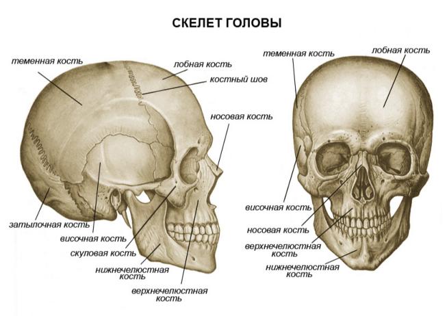
Facial bones- their main function is to participate in chewing food.
Bones of the brain skull- The cranium consists of eight flat bones that protect the brain, connected motionlessly.
Ribs- these are the bones that, together with the sternum, form chest, a necessary element of protection of the internal organs located in it.
Spinal column- the axis, or support of our body, consisting of 33 or 34 vertebrae, it houses the spinal cord.
Femur- the longest bone in the human body. Allows a variety of leg movements due to its connection to the kneecap.
Foot bones- a group of 26 bones, among which the largest, the calcaneus, forms the heel. The most tall man There was an American in the world whose height was 2.72 m. At the time of his death, in 1940, when he was 22 years old, he was still growing. The shortest person was a 19-year-old Dutch woman: her height was only 59 cm, she died in 1895. The longest bones known to exist are those of Brachiosaurus, a dinosaur whose remains were found in Colorado (USA). Its shoulder blades reached a length of 2.4 m, and some ribs exceeded 3 m. Among modern living creatures, the tallest animal on Earth is the giraffe, its height can reach 6 m. The long neck, more than 2 meters, necessary for the giraffe to feed on tree branches, includes only seven cervical vertebrae, the same as a mouse. Perhaps the smallest are the temporal bones of the hummingbird - a bird whose length does not exceed 2-3 cm, but which has muscles on its wings that allow it to make up to 90 beats per second. Hummingbirds can hover in the air while feeding on flower nectar, and can even fly backwards. Muscles, of which there are more than 400, cover the skeleton and, together with the bones and their joints, make movement possible, but some of them, for example the muscles of the veins and arteries that provide the flow of blood pumped by the heart, perform functions not related to the locomotor system.
Year after year, more and more aspects of life activity are revealed, over which the brain extends its supreme influence: metabolism, control of physical and chemical processes in the blood, hematopoiesis, the fight against infectious principles, etc., etc. How endlessly This is far from those inconspicuous fibers, which had barely begun to separate from the surrounding tissue, along which the primitive electrochemical excitatory impulse made its way! In higher, neokinetic animals, including us, movements are guided by sensations, controlled and directed by them. In the lower classes, on the contrary, sensations are served and provided with the help of movements. Movements; seemingly haphazard and confused, they go ahead of sensations, grab and catch them anywhere. This mechanism of active, active “sensation” has been preserved among us, with the exception of unsystematic nature, in the work of our highest sense organs, vision and touch, where the circulation of the “reflex ring” is woven into a completely inseparable and very complex structure. In subsequent essays we will have several more cases of seeing with what care our central nervous system generally preserves the most ancient mechanisms, which would seem to have long been outdated and should be archived. This crude ancient mechanism of sensation, which operated in the most distant times, long before sensory corrections, was again revived in an improved and refined form and, merging in its work with these corrections, ensured the functioning of our most highly developed sense organs.
Facial muscles- allow us to take on different facial expressions: laughter, anger, etc.
Biceps brachii- together with its antagonist - the triceps brachii muscle - ensures flexion and extension of the forearm.
External obliques- allow air to be expelled from the lungs during contraction. They perform the opposite work of the diaphragm, which is not visible here, since it is located inside the abdominal cavity.
Quadriceps femoris- as in the case with upper limbs, the quadriceps femoris muscle also has an antagonist muscle - biceps muscle hips. Both flex and extend the hip.
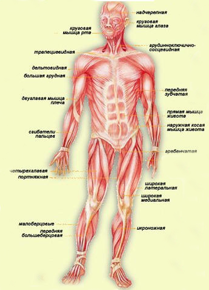
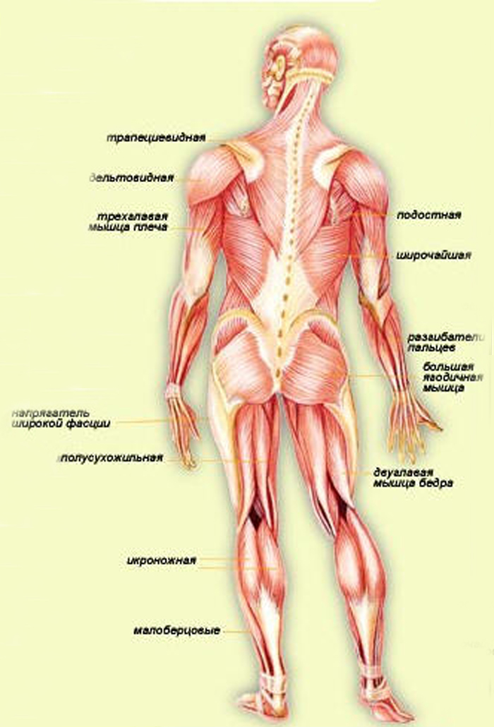
Diagram of the human musculoskeletal system (front view)
Regarding sensory corrections, it should also be added that the necessary need for them, revealed in higher animals, served as a new and very powerful incentive for the further development of the brain. As we will show later, this need mainly contributed to the development of the so-called sensory fields, that is, entire complex casts from the sensations of a wide variety of senses, casts that guide the movements of an animal or a person and help organize these movements in space.
Limb development
The second innovation, which naturally followed the consolidation of the neokinetic system with its articulated levers and striated muscles, was the development of animal limbs. Lower, non-skeletal organisms did not have limbs; at best, instead of them, “false limbs” (pseudopodia) like rays sometimes appeared starfish or the “legs” of a snail, which, in fact, is the bottom of its body. And vertebrates did not develop real limbs right away.
The limbs were a very deep, fundamental innovation. They appeared at a time when the ancient motivations for the segmented structure of the body had largely fizzled out and the development of the limbs began, as it were, stepping over the ruins of this ancient principle of structure, which was still preserved on the oldest part of the body - the torso. Therefore, firstly, the limbs themselves no longer show any traces of segmentation - this can be seen at least in the way their muscles are supplied with motor nerves. Secondly, we need to point out here one circumstance that is much more important for our presentation. The consistent development of neokinetics in vertebrates, followed by greater motor synergies for movement in space (locomotion), and finally, limbs as improved tools for such movement, led to a corresponding enrichment of the central nervous system with devices necessary to serve all these evolutionary innovations. Comparative anatomy of the animal brain shows that this entire series of innovations, more than any of the previous steps of development, contributed to real centralization in the brain, the appearance in it of the first formations that without reservation deserve the name brain. The oldest part of the central nervous system of vertebrates, the spinal cord, is still entirely of the segmented type of structure. The new nuclei of the brain, developed during the “fish” period of vertebrate evolution and finally formed in the first animal with legs - the frog, are already completely suprasegmental. Their nerve conductors control the entire spinal cord as a whole, and in particular all the limbs. It is even more important to note the fact that the activity of this supreme brain, which controls the movements of the limbs and locomotion (we will designate it in subsequent essays as level B), occurs in amphibians completely according to the laws of the neokinetic system: with relatively high-voltage and fast-moving electrical signals, with obedience to the law “all or nothing”, etc. The more ancient centers of the brain, behind which amphibians retained control of the body (level A according to our designations), work to a large extent according to ancient motor laws: with low-voltage, slow impulses, with a high degree of participation they contain ancient, chemical signal transmission, etc. The remarkable thing here is that even we, people, owners of a brain that is more different from the brain of a frog than a multi-story palace from a savage’s shack, even in our brain there are separate form level B and level A, with decent clarity dividing between themselves the control of the limbs and cervical-trunk muscles, and even in our country the ancient, segmental, trunk level A to a large extent continues to work according to the same ancient motor laws. We will cover the issue of levels more fully in the next two essays.
Movement enrichment
All subsequent development of movements in vertebrates is a continuous enrichment of the motor means and capabilities of animals from class to class and from “year” to “year” of our chronological table of their evolution. This enrichment does not occur without a reason and not as a result of some mysterious internal “spring” inherent in animals, which impels them to continuous improvement. No, the same tough and ruthless, purely external reason always leads to the enrichment of motor resources: competition and the struggle for life. Animals become cramped from continuous reproduction. They lack food. Predatory breeds are being developed that prefer to allow other animals to find suitable nutritional material for themselves and capture it in a ready-made, “semi-finished” form, devouring these weaker animals. These latter develop means of self-defense: quick legs, protective coloring, armor coverings, horns and hooves, etc. Without such means of protection, they are first of all devoured by predators, who, without knowing it, contribute to the improvement of the breeds they are pursuing. In fact, the greatest chances of surviving extermination and producing offspring similar to themselves for a long time are those individuals who, perhaps even by chance, are better protected. And the most reliable self-defense is still rich and perfect motor capabilities. The same law of competition hits predators with the other end of the stick: those who are insufficiently agile, cunning and toothy among them risk dying of hunger, not being able to capture edible living creatures that have contrived in self-defense.
Movements are enriched in this way primarily in their strength, speed, accuracy and endurance. But this enrichment is almost only quantitative. More important are the other two sides of the movements, which are becoming more and more improved. Firstly, the motor tasks that an animal has to solve are becoming more and more complex and at the same time more and more diverse. The entire list of movements of a fish consists almost entirely of its main locomotion - swimming and some pair of simple hunting movements in addition. In one of the most poorly developed fish, the shark, its entire hunt consists in the fact that it swims under its prey, turns its belly up (this is more capable for it) and opens its mouth. In addition to swimming, an amphibian animal can also crawl, jump, and make sounds. The snake already knows how to hide in ambush. And how complex and full of variety, in comparison with all this, are even the chain hunting actions of a mammalian predator! There are the cunning of a fox, the sensitive search of a hunting dog, and the insidious ambush of a tiger aiming at prey that is not easy for him. In the coming lines we will trace in more detail this side of the movements, the complication of the problems they solve.
Secondly, the number of unforeseen, non-routine tasks that the animal has to solve right there, “on the fly,” is increasing. As we have already seen in the introductory essay, this is precisely where the greatest demand for dexterity takes place. In the motor life of an animal, there are becoming relatively fewer and fewer standard, always identical movements that can be performed automatically, without delving into anything or adapting to anything. One could assume that, for example, locomotion, movement through space, is an example of similar, eternally patterned movements. This is far from true. When a fish swims inside a boundless aquatic environment that is homogeneous in all directions, there really isn’t much reason for diversity. But moving on land is a completely different matter, which in nature does not take place on treadmills. There are ditches, gullies, swamp hummocks, and impassable thickets; here are safe paths along which you can trot, and a forest full of secret enemies, where you need to sneak silently, alerting all your telereceptors, etc., etc. What can we say about more complex motor acts, completely inaccessible fish and overflowing with the life of a highly developed mammal? The many times intensified struggle for life makes his existence full of surprises, and surprises require the ability to immediately, at the cost of a hundredth of a second, make the correct motor decision and accurately, deftly implement it. We will see further that this non-stop increase in the number of unlearned movements and actions is based on the same non-stop development of completely new, higher parts of the brain, mainly the so-called cerebral cortex.
The first rudiments of the cerebral cortex appear already in higher reptiles, but only in higher vertebrates - in mammals - does it take decisive dominance and continuously develop further and further. It is the cerebral cortex that is the organ of the brain that has the unlimited ability to absorb the personal life experience of an animal, remember it, meaningfully master it and create on its basis one-time solutions to new, never encountered problems. In terms of mental activity, this ability is intelligence, ingenuity, intelligence; in terms of motor acts, we call this same ability dexterity. It is not for nothing that they often say about a person endowed with pronounced dexterity: “What smart movements he has! What smart hands.” The brain matures in a human baby, floor by floor, in the same order in which they arose in the animal world. The baby is born from just - the pallidum level B is just finishing its development - the "ceiling" level of amphibians. Therefore, the child is not able to make any movements that would go beyond the meager list of this level. The matter is further complicated by the fact that the more ancient and lower located level A , which will be discussed below and which controls the movements and positions of the neck and torso, does not have time to mature and become operational by the time of birth.Because of this, the result is, first of all, that the newborn cannot control the main support of the whole body - the torso and neck, holding his head, and therefore unable to use his “dynamic supports" - his limbs. His torso lies helplessly on his back, heavy and motionless, and all four legs can only make random, idle kicking movements in all directions. And besides this, there is another complication: level-floor B, as already mentioned, has access for its impulses to the motor cells of the spinal cord, and through them to the muscles only by “transit”, through the nuclei of the underlying level A. Therefore, it and he himself is forced to wait in inaction until Level A finally matures and begins to pass through its motor impulses. This deprives the child of the synergy that level B brings with it - coordinated integral movements of the limbs, and even more so collaboration all limbs. Practically speaking, during the first two to three months after birth, there is no motor coordination of any kind. Only by the end of the first quarter of life do correct joint eye movements, turns from back to stomach, etc. begin to be organized. Around the end of the first half of the year, more or less simultaneously, the lowest level A, which gives the baby a coherent and strengthened torso, and level striatum (CI), which gives him the ability to sit, stand on his legs, stand, then crawl on all fours (again, a biogenetic memory of our four-legged ancestors!) and, finally, walk and run. The pyramidal system of the cortex (PCS) is delayed even more. The sensitive parts of the cortex begin to work much earlier: the child begins to recognize what he sees, and understand the words addressed to him, and find an understanding of taste and gastronomic sensations. The PDS begins to gradually manifest itself during the second half of the year, following the striatum system. This is reflected in the fact that the child learns to grasp what he sees in front of him, put and rearrange things, point with his finger, etc. The first monosyllabic meaningful speech sounds, usually commanding and pleading (like " give!"). The movements of the arms are still very inaccurate, the child often and roughly misses, but until this time he had not even attempted to make movements such as grasping or throwing. He had nothing to do with them! The difference between infants after and before six months in relation to these movements is approximately the same order as the difference between the owner of a bicycle, who still barely knows how to ride it, and a person who does not have a bicycle at all. So, the intensification of the struggle for existence gradually accumulated more and more significant amounts of homogeneous motor tasks, which were so far beyond the capabilities of animals. The need to cope with them matured over time with increasing inevitability. The animal had to satisfy these increasingly complex motor needs at all costs if it did not want to die. And on the way to such satisfaction there was one obstacle, the main and main one: the need to master new sensory corrections.