The shoulder muscles involved in the movement of the elbow joint, in turn, are divided into two groups. The anterior group consists of flexor muscles: the brachialis and biceps shoulder The posterior group includes extensor muscles: the triceps brachii and the anconeus muscle.
Brachialis muscle starts from the lower half of the front surface humerus and from the intermuscular septa of the shoulder, and is attached to the tuberosity of the ulna and its coronoid process. The brachialis muscle is covered in front by the biceps brachii muscle. The function of the radialis muscle is to participate in flexion of the forearm.
Biceps brachii has two heads, starting on the scapula from the supraglenoid tubercle (long head) and from the coracoid process (short head). The muscle is attached on the forearm to the tuberosity of the radius and to the fascia of the forearm. It belongs to the biarticular muscles. In relation to the shoulder joint, the biceps brachii muscle is a flexor of the shoulder, and in relation to the elbow joint it is a flexor and supinator of the forearm.
Since the two heads of the biceps brachii muscle, long and short, are attached to the scapula at some distance from each other, their functions in relation to the movement of the shoulder are not the same: the long head flexes and abducts the shoulder, the short head flexes and adducts it.
In relation to the forearm, the biceps brachii muscle is a powerful flexor, since it has a much larger shoulder of force than the brachialis muscle, and, in addition, it has an instep support, much stronger than the actual instep support of the forearm. The supinator function of the biceps muscle decreases somewhat due to the fact that the muscle passes into the fascia of the forearm with its aponeurosis.
The biceps brachii muscle is located on its anterior surface directly under the skin and its own fascia; the muscle is easily palpable both in its muscular part and in the tendon part, at the point of attachment to the radius. The tendon of this muscle is especially noticeable under the skin when the forearm is bent. Under the outer and inner edges of the biceps brachii muscle, the medial and lateral brachial grooves are clearly visible.
Triceps brachii located on the back of the shoulder, has three heads and is a biarticular muscle. It is involved in the movements of both the shoulder and forearm, causing extension and adduction in the shoulder joint and extension in the elbow.
The long head of the triceps muscle starts from the subarticular tubercle of the scapula, the amedial and lateral heads - from the posterior surface of the humerus (medial - below, and lateral - above the groove of the radial nerve) and from the internal and external intermuscular septa. All three heads come together to one tendon, which, ending on the forearm, is attached to the olecranon process of the ulna.
This large muscle lies superficially under the skin. Compared to its antagonists, the shoulder and forearm flexors, it is weaker.
Between the medial and lateral heads of the triceps brachii muscle, on the one hand, and humerus, on the other, there is the brachiomuscular canal; the radial nerve and the deep brachial artery pass through it.
Elbow muscle originates from the lateral epicondyle of the humerus and the radial collateral ligament, as well as from the fascia; it is attached to the upper part of the posterior surface and partly to the olecranon process of the ulna in its upper quarter. The function of the muscle is to extend the forearm.
The fascia of the forearm is highly developed, especially on the back of the forearm. In the form of a dense case, it covers the muscles of the forearm and separates them by intermuscular septa. Posteriorly, the fascia of the forearm is attached to the olecranon process and the posterior edge of the ulna. Distally it passes into the fascia of the palm and dorsum of the hand. At the border with the hand it forms thickenings, which are called on the dorsal surface the extensor retinaculum, and on the palmar surface the flexor retinaculum, which strengthen the tendons of the muscles running from the forearm to the hand and to the fingers, creating the most favorable conditions for the manifestation of muscle strength.
Today I’ll tell you how one of the most iconic muscles works - the biceps brachii muscle (BMB), in common parlance the biceps or “ jar" As a rule, in the gyms, everyone is mainly engaged in pumping cups..)), no matter how you look, they do all the barbell lifts for biceps, as if there were no other muscle groups. If among intellectual people they meet by their intelligence, then among athletes they meet by their banks..)). Perversion, in a word.
Start - Attachment.
The DMP has two heads, long And short. The long head starts from the supraglenoid tubercle of the scapula, and the short head starts from the coracoid process of the scapula. The muscle is attached to the tuberosity of the radius and to the fascia of the forearm (tendon).
Function.
The DMP is biarticular. It flexes the shoulder and fixes the head of the humerus in this joint; in relation to the elbow joint, it is a flexor and supinator of the forearm. Since the heads of the shoulder blade begin on the scapula at some distance from each other, their functions in relation to the movement of the shoulder are different: the long one flexes and abducts the shoulder, the short one flexes and adducts it. In relation to the forearm, the DMP is a flexor, as it has a significant leverage.
Characteristic.
The ASD is located on the front of the shoulder, just under the skin. According to its target action it is agonist, if it helps, is synergist.
Exercises for the biceps brachii muscle.
Dumbbell flyes lying on a horizontal bench. Synergist.
Bringing your arms together in a “crossover” frame. Synergist.
Raising dumbbells forward with outstretched arms. Synergist.
Arm curls with a barbell (any weight: free weight, machine handle) in the hands while standing (sitting), Agonist. Any variations (on a Scott bench, one arm, etc.).
All types of pull-ups to the bar. The end of the humerus closest to the body is affected by the weight of the body, which creates rotation, extending the humerus at the elbow. Accordingly, the DMP, together with nearby muscles, resists and bends the shoulder in relation to the forearm.
And to sum up, I can say, as I always repeat, any muscle on the human body works with any complex movements. What is worth striving for.
Dear readers of my blog!
If you want to get your body in order. Don't waste your time looking for a miracle pill, don't miss the opportunity to give yourself physical activity without leaving home. Especially if you can’t go to the gym. Either the child is nearby, or there is simply no gym nearby.
I offer you classes within walking distance. All you need is the Internet and a place in a 2x2 house. Further, everything looks like training under the guidance of a trainer in the gym. Only, it’s much more economical, both in terms of money and time. And most importantly, no one stares at you or interferes.
Video chat classes look like this:
You can see what the process of launching classes and the cost looks like by clicking on the link below, but first take a look at those who are no longer wasting time:
You can easily contact me via Viber – 89258591633 or Telegram – @samotoyloff. You can also find my coordinates in the lower right corner of the site ( clapping hands).
Don't waste time, join those already practicing. The process is no different from what is happening in the hall. I see you the same way, I control you the same way, I tell you what and how to do.
Proximal attachment. Long head: supraglenoid tuberosity of the scapula. Short head: coracoid process of the scapula.
Distal attachment. Tuberosity of the radius.
Function. Flexes the forearm at the elbow joint; promotes flexion of the shoulder at the shoulder joint. Moves the shoulder away from the body and at the same time turns it inward.
Palpation. For localization it is necessary to identify the following structures:
. Intertubercular groove of the humerus - Locate the greater and lesser tuberosities of the humerus, lying just distal to the lateral surface of the acromion. (It is most convenient to palpate them with the hand turned outward.) The groove lies medial to the greater tuberosity and lateral to the lesser tuberosity. The tendon of the long head of the biceps brachii muscle runs along the interbud groove.
The coracoid process of the scapula extends from the superior edge of the scapula between the neck and notch of the scapula. Find the most concave surface of the lateral clavicle; move the palpating hand distally approximately 2.5 cm into the deltopectoral triangle. When you press posterolaterally, you will feel a bony protrusion - the coracoid process. This area can be very sensitive.
The powerful biceps brachii muscle can be palpated along its entire length. Flex your shoulder 15 to 45 degrees to locate the tendon that attaches to the radial tuberosity. Palpate the biceps muscle upward. The long head can be palpated by following its tendon, which runs along the intertubercular groove; Palpation of the tendon and groove is easier when the shoulder is rotated outward. The short head is palpated medially in the direction of its attachment to the coracoid process of the scapula.

Pain pattern. Superficial aching pain on the front of the shoulder and shoulder joint with some limitation of mobility.
Causal or supporting factors.
Prolonged elbow flexion; Chronic or acute sprains from playing sports or lifting heavy objects.
Satellite trigger points. Brachialis, triceps brachii, muscle that rotates the forearm outward (supinator).
Affected organ system. Respiratory system.
Associated zones, meridians and points.
Ventral zone. Hand meridian lung tai yin, hand meridian pericardium jue yin. Ш 3, 4, 5; PC 2, 3.
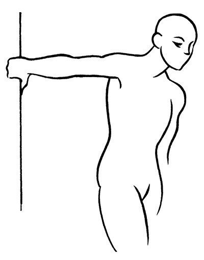
Stretching exercise. Grab the door frame with your affected hand. The palm should be at shoulder level, the elbow straight, and thumb directed downwards. Rotate your torso away from your shoulder without allowing your arm joints to bend. Fix the pose until the count is 15-20.
Strengthening exercise. Stand straight, arms at your sides, palms facing inward. Bend your forearms without moving your elbows away from your body. Stretch your palms towards your shoulder joints. Slowly return to the starting position. Perform bending on a count of 2, return to the starting position on a count of 4.
Now stand in the same way, but turn your palms outward. Bend your forearms without moving your elbows away from your body. Stretch your palms towards your shoulder joints. Slowly return to the starting position. Perform bending on a count of 2, return to the starting position on a count of 4.
Repeat the exercise 8-10 times, increasing the number of repetitions as your strength increases. You can use dumbbells to increase the load.
D. Finando, C. Finando
The movement of the joints of the limbs is carried out due to the work of the muscles located on them. They consist of special fibers arranged in bundles. In the human body there are about 400 muscles that, under the influence of impulses from the central nervous system, are able to change the position of the body.
Types of muscle tissue
Movement is impossible without the participation of muscles. The total mass of such tissues in the adult human body is around 30-40%. In newborn babies it is about 20%, in older people it decreases to 25-30%. But if a person remains active even at an advanced age, then muscle mass does not decrease, it remains at the same level.
Experts distinguish different muscle groups depending on their location, functions, and direction of fibers. The following types of muscles are distinguished by location:
Superficial (located under the skin);
Deep;
Lateral;
Medial;
External;
Internal.
According to their shape, experts distinguish fusiform, square, triangular, circular, and ribbon-shaped muscles. According to the number of heads, they can be two-headed, three-headed and four-headed. Depending on the direction of the bundles, they are single-pinnate, bi-pinnate or multi-pinnate.
Muscles are also classified according to their functions. Some provide extension of the limbs, others - flexion. Separately, the rotator-lifters, compressors (sphincter), abductor and adductor muscles are distinguished.
For example, the biceps brachialis is fusiform. It is attached to a bone that acts as a lever and allows the arm to flex at the elbow.
Biceps shoulder
 The biceps belongs to the anterior group of muscles. It is located on the front of the humerus. This long fusiform muscle has two heads. One of them is long, the second is short. They walk side by side, heading from top to bottom. At the level of the middle part of the shoulder they are connected into a spindle-shaped abdomen.
Moreover, if the semitendinosus and semimembranosus muscles are located medially, then the biceps muscles occupy a lateral position. The femoral biceps extends along the entire length of the femur and has a fusiform shape. It is adjacent to the vastus lateralis muscle. The lateral wall of the popliteal fossa is formed by the biceps femoris muscle. It can perform its functions thanks to its special structure and attachment to another bone.
It starts with two heads - long and short. At the beginning of the first of them there is a short thick tendon. It is located on the surface of the sacrotuberous ligament and the ischial tuberosity. The muscle does not go straight down, but obliquely in the lateral direction. The lower end is attached to the lower leg.
The short head of the biceps femoris muscle originates from the posterior surface of the femur. Its lower edge ends at the tibia (tibia).
Both heads connect at the border between the lower and middle parts of the femur. They pass into one common tendon. It runs from the back of the knee joint. The specified tendon is attached to (its head) and the outer part lateral condyle related to tibia. Partially its fibers are woven into the fascia of the lower leg.
Functions of the biceps
 Depending on what work the muscles can perform, they are divided into flexors and extensors. These are opposing muscle groups that provide movement of the limbs. The main flexor muscle of the upper arm is the biceps brachii.
It performs the following functions:
Flexes the arm at the elbow joint;
Provides the ability to rotate the brush outward;
Tenses the arm in the elbow joint.
The main flexor of the upper leg is the biceps femoris muscle.
Functions of the leg biceps:
Hip extension and adduction;
Straightening the body after bending;
Flexion of the lower leg at the knee joint;
Rotating the shin outward, bent at the knee joint;
Maintaining balance.
It is worth noting that problems with the biceps, such as insufficient strength or flexibility, lead to back pain, problems with the knee joints, and poor posture.
Extension of the upper limbs is achieved through the activity of the triceps, located along the humerus. The biceps and triceps muscles can contract together or alternately. The movement of the leg in the knee joint is facilitated by the coordinated work of the biceps and quadriceps muscles.
Leg extensor
Most big muscle V human body is the quadriceps. It is located on the front surface of the thigh and provides the ability to straighten lower limb in the knee area. It is also responsible for flexing the hip - bringing this part of the leg closer to the stomach.
The quadriceps consists of four bundles. Each of them is considered a separate muscle, which has its own name. Separately, experts distinguish the rectus, vastus lateralis, medialis and intermediate muscles.
All of them are attached to the patella. But each of them has its own functions. For example, the straight line is responsible for hip flexion and extension knee joints. But the intermediate, medial and lateral are necessary for straightening the lower leg.
Providing traffic
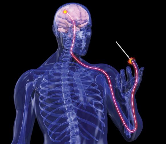
 Without the coordinated action of the muscles, it would be impossible to bend or straighten the limbs. Voluntary movements of the biceps muscle are regulated by the spinal cord. This becomes possible due to the alternation of the processes of inhibition and excitation that take place in the spinal cord. The muscles responsible for flexion and extension of the limbs can simultaneously be in a relaxed state. This happens, for example, while the arm hangs relaxed along the body.
Their contraction occurs due to the activity of spinal cord neurons responsible for movement. The relaxation process is associated with inhibition of these cells of the nervous system. If a person holds a dumbbell in a straight outstretched arm, the biceps and triceps muscles will work simultaneously.
When nerve impulses arrive, the muscle receives a certain command, depending on this, the process of relaxation or tension of the muscle begins. When contracted, it acts on the bone to which it is attached like a lever.
The principle of muscle functioning
 Contracting any muscle group is work that requires energy to perform. Its source may be products released during the decomposition and oxidation of various organic substances. In the exercise of muscles, they are exposed to various chemical reactions. The result of such interaction is the release of energy. This is accompanied by the formation of cleavage products - carbon dioxide and water.
When too much stress is placed on the muscles, a natural process of fatigue begins. This is accompanied by a decrease in their performance. After rest, everything is restored.
It was noted that when performing rhythmic exercises, fatigue sets in somewhat later. After all, in the intervals between contractions, the muscles partially have time to rest and recover. But performance is also affected by the intensity of the load. The higher it is, the faster fatigue will set in.
Work of the central nervous system
When making this or that movement, a person does not think about it. It does everything automatically. But at the same time, each motor act for the nervous system is a complex process, the implementation of which requires the use of its various levels. All active movements are controlled by the brain. They are called voluntary or conscious.
Before muscle contraction begins, the cerebral cortex receives information through special channels about the state of the joint-muscle fibers. She assesses how ready they are for the load. Therefore, it cannot be said that voluntary movements of the biceps muscle are regulated exclusively by the spinal cord. After all, the cortex controls the strength, sequence and duration of each contraction.
The motor centers are located in the frontal cortex of the brain. It is in the anterior sections that all signals are integrated. After this, a model of future movement is formed.
In order for voluntary muscle contraction to occur, impulses generated in the cortical part of the brain must reach the corresponding muscles. They pass along a special path, which experts call the cortical-muscular one. Voluntary movements of the biceps muscle are regulated by central and peripheral neurons. The first of them are the bodies of pyramidal cells with axons. The second are the cells of the spinal cord.
Neurons connect the part of the cerebral cortex responsible for movement, special parts of the spinal cord and brain stem. This entire complex is called the pyramidal system.
Possible problems
 It happens when some part of the corticomuscular pathway is affected. In this case, the muscles do not receive the signal coming from the cerebral cortex. Their voluntary movements become impossible.
For example, with partial damage, the biceps muscle cannot perform its functions in full. Depending on the location of the lesion, problems can arise in different parts of the body. Partial damage usually leads to paresis.
With such injuries, voluntary movements of the biceps muscle are also regulated by the brain and spinal cord. But due to disruption of connections, disruption and limitation of the intensity and strength of contractions is possible. The problems may be central or peripheral depending on which neurons are affected.
The examination of such patients must be thorough. During its implementation, it is important to determine not only how motor function has been preserved, but also to check for the presence of muscle atrophy. They also look at whether there is deformation chest, spine, whether small muscle twitches are present.
But it is worth noting that it is not always only because of problems with the cortico-muscular pathway that movements become impossible. For example, with pathologies of the articular apparatus, disorders of proprioceptive sensitivity, the biceps brachii muscle may stop working. It will not perform its functions fully even if scar changes have appeared in the muscles. Therefore, it is important to determine the reason why muscle contractions have become impossible. For these purposes, they examine how the patient can perform active and passive movements, and evaluate his reflexes.
Many people know about a muscle such as the biceps (biceps muscle), but few people understand the features of its structure and its true name. It is extremely difficult to pump up your arms without this information, so it is advisable to familiarize yourself with all the anatomical details before drawing up a training plan. In this case, you can achieve results in a shorter time.
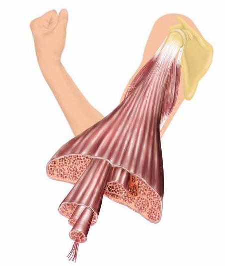
The biceps brachii muscle is attached to the bones of the scapula by a tendon. Muscle tissue gets its name from the two heads that can be seen in this image:
The picture clearly shows what parts the biceps brachii muscle consists of, namely:
- Short head of biceps. This part of the biceps brachii begins with the coracoid process on the outside of the shoulder blade. From here the muscle runs along the inner surface of the bone to the long head. The short half of the biceps does not have an oblong tendon, but it contains more muscle tissue;
- Long head of biceps. It is localized on the lateral surface upper limb and begins its path from the protrusion in the area of the scapula, which is located directly above the recess of the shoulder joint. This place is called the supraglenoid tubercle. The long head has a fairly pronounced tendon, but at the same time a short section of muscle tissue.
If you look from above at the structure of the biceps of the arm, you will notice that both heads intertwine with each other as they approach the elbow joint, creating a kind of abdomen. It is attached to the elbow using the biceps tendon. Together, both heads create a powerful flexor, that is, a flexor.
Function
By studying the structural features of the biceps brachii muscle, you can understand what its main functions are. According to its anatomy, the biceps is a flexor of the limb at the elbow joint and allows you to rotate (supinate) the hand. The long head of the muscle comes into action when the shoulder muscle tissue contracts, for example, when raising the arms upward.
To fully stretch the long part of the biceps, your elbows will need to be pulled back. If you need to load the short head of the biceps, they need to be moved slightly forward from the body. This nuance is useful for novice bodybuilders, since certain hand positions affect the pumping of problematic muscle tissues. That is why any athlete should study the functions of the biceps before pumping up their arms. 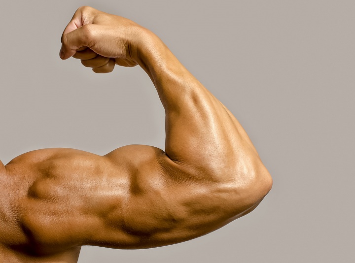
Stress points
The biceps brachii are constantly loaded during training and excessive strain can create stress points. For example, this can happen during a hill climb or after a barbell bench press. In bodybuilders, the main reason for the appearance of such spots is carrying a lot of weight on the elbows or outstretched limbs. However ordinary people are also not immune from them. After all, any activity accompanied by severe overload can cause pain and weakness in certain areas of the arm, which are symptoms of stress points.
You can find out about their presence by pain in the biceps muscle area. Sometimes the discomfort is localized on the front surface of the shoulder. At the same time, people pay attention to the restrictions that have arisen that were not there before. For example, weakness when straightening the arm or the appearance of pain when palpating the tendon that attaches the biceps to the elbow.
To identify such points, you should place your limb on a flat surface in front of you. First you need to bend it slightly at the elbow joint. Then, using palpation, you should look for points of tension.
You need to start palpation from the ulnar fossa and gradually move along the tendon to the belly of the biceps.
You should not just poke with your fingers, but move smoothly, massaging the outer and inner sides of the muscle tissue. As you move, you can feel the seals and there are often stress points near them. They are usually localized 1/3 of the way along the biceps muscle. If such places are found, they need to be massaged until the discomfort decreases.
Biceps muscle pain
 The biceps brachii muscle usually endures loads, but sometimes pain of a different nature occurs in it. In such a situation, you need to know the reasons why it may occur:

If suspicious symptoms are detected in the biceps muscle, you should consult a therapist. To conduct an examination, he can send the patient to a physiotherapist, traumatologist, rheumatologist, etc. After identifying the main cause of pain, an appropriate course of therapy is prescribed.
The biceps muscle is known to most people as the biceps. Knowing its features, you can quickly and effectively pump your arms and avoid injuries associated with excessive overload. However, this muscle is under constant tension, so it is necessary to ensure that strange symptoms do not occur. If they are detected, you should immediately contact a specialist.